Cherry Eye In Dogs: Comprehensive Guide
Learn about cherry eye in dogs, why it happens, how to spot it, and what treatments keep your dog's eyes comfortable and healthy.

Image: HearthJunction Design Team
Understanding Cherry Eye in Dogs
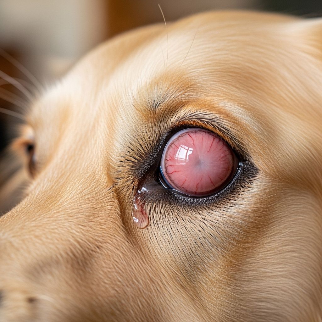
Cherry eye is a term commonly used by veterinarians and dog owners to describe a distinctive and sometimes alarming red or pink mass that appears in the inner corner of a dog’s eye. While it may seem frightening at first glance, cherry eye is a relatively well-understood condition, officially known as a prolapse of the third eyelid gland. This guide provides a comprehensive overview of cherry eye, including its causes, diagnosis, symptoms, risk factors, and treatment options.
What Is Cherry Eye?
Unlike humans, who have just two eyelids, dogs possess a third eyelid, called the nictitating membrane. This third eyelid is not usually visible, but it plays a critical role in protecting the eye and maintaining proper lubrication by dispersing tears across the eye’s surface. Attached to this membrane is a tear gland responsible for producing a significant portion—up to 50%—of the watery component of tears a dog needs for healthy eyes.
Cherry eye occurs when the connective tissue holding the third eyelid gland in place weakens, causing the gland to pop or prolapse out of its normal position. When this happens, the gland protrudes and appears as a pink or reddish lump in the inner corner of the eye. It is commonly referred to as “cherry eye” because of its round, cherry-like appearance.
What Causes Cherry Eye?
The root causes of cherry eye are not fully understood, but the primary culprit is thought to be a congenital weakness in the connective tissues that anchor the third eyelid gland. This hereditary predisposition means some dog breeds are more susceptible than others. While injury or trauma may also trigger the gland’s prolapse, it is most often seen in young dogs, especially those under two years of age.
- Genetics: Certain breeds have an increased risk due to weak connective tissues.
- Age: The condition most frequently affects dogs younger than two, with a significant majority (around 75%) of cases occurring in those under one year old.
- Trauma: Injury to the eye area may contribute, although this is less common.
- Breed Predisposition: See common breeds at risk below.
Breeds Most at Risk
While cherry eye can develop in any dog, some breeds are particularly predisposed due to genetics:
- Bulldogs (English and French)
- Cocker Spaniels
- Lhasa Apsos
- Beagles
- Boston Terriers
- Pekingese
- Shih Tzus
- Bloodhounds
- Saint Bernards
- Great Danes
These breeds are among those most commonly seen with cherry eye, but the condition is not exclusive to them and can affect any dog.
Signs and Symptoms of Cherry Eye
Cherry eye is usually very noticeable to pet owners. Its most obvious symptom is the sudden appearance of a red, pink, or reddish mass in the corner of a dog’s eye, near the nose or muzzle. This abnormal tissue is the prolapsed gland of the third eyelid.
- Round, red or pink mass protruding from the inner corner of the eye
- Inflammation and mild swelling around the affected area
- Watery eyes or increased tear production
- Occasional discharge from the eye
- Possible squinting or blinking more than usual
- Pawing at the eye or face (a sign of discomfort)
Although cherry eye itself is not generally painful, it can cause irritation, discomfort, or lead the dog to rub or scratch at the eye, increasing the risk of secondary injury or infection.
Diagnosing Cherry Eye in Dogs
Diagnosis of cherry eye is straightforward and typically based on a visual examination by a veterinarian. The appearance of a pink or red mass in the inner eye corner is distinctive enough for most vets to make a confident diagnosis.
However, a thorough ophthalmic exam is always performed to assess the extent of the issue and rule out other eye disorders. This examination may include:
- Testing vision and ocular reflexes
- Measuring intraocular (eye) pressure
- Applying a fluorescent dye to detect scratches or ulcers on the eye surface
- Measuring tear production using the Schirmer tear test (placing a paper strip on the lower eyelid to assess tear output)
In rare cases, further diagnostic testing may be required if other eye diseases are suspected.
Is Cherry Eye Dangerous?
While cherry eye is not typically painful, it is not a condition that should be ignored. The exposed gland is vulnerable to drying out, trauma, and infection. Over time, untreated cherry eye can compromise tear production and potentially lead to serious eye conditions, such as keratoconjunctivitis sicca or “dry eye,” which can permanently impact a dog’s vision.
Seeking prompt veterinary care is essential to:
- Relieve discomfort
- Protect the eye from further injury or infection
- Preserve long-term eye health and tear production
Treatment Options for Cherry Eye
Cherry eye will not resolve on its own and requires veterinary intervention. The primary goal is to restore the gland to its normal position and maintain adequate tear production. Surgery is the treatment of choice.
Why Is Surgery Necessary?
In the past, some veterinarians would surgically remove the prolapsed gland. However, because this gland produces up to half of the tears needed to keep the eye healthy, removal often resulted in chronic dry eye and other complications. Today, surgical repositioning—not removal—is considered the gold standard, unless the gland is nonfunctional due to cancer or severe injury.
How Is Cherry Eye Surgery Performed?
A few different surgical techniques are used to correct cherry eye, but the two most common are:
- Mucosal Pocket Technique: The surgeon creates a small pouch or pocket in the corner of the eye, tucks the prolapsed gland back into this new pocket, and secures it using absorbable stitches.
- Orbital Rim Technique: The gland is anchored to the bone around the eye with permanent sutures for enhanced stability. The choice of technique depends on the individual dog and the surgeon’s preference.
| Technique | Summary | Pros | Cons |
|---|---|---|---|
| Mucosal Pocket | Creates a pocket in the third eyelid for the gland | Minimally invasive, quick recovery | Potential for recurrence |
| Orbital Rim | Anchors gland to surrounding bone with stitches | Secure, less likely to recur | More invasive, longer healing |
What to Expect During and After Surgery
- General anesthesia is required for all cherry eye procedures to ensure the dog’s comfort and safety.
- After surgery, dogs may need
- Protective collars (E-collars) to prevent scratching at the stitches
- Antibiotic or anti-inflammatory eye drops to reduce swelling and prevent infection
- Pain relief medication, as prescribed by the vet
- Most dogs recover within a few weeks and regain normal function of the third eyelid gland.
What If the Gland Prolapses Again?
Unfortunately, recurrence is possible. Statistics show that about 5–20% of patients may experience the gland slipping out of place again. Repeat surgery is sometimes necessary.
Gland Removal: When Is It Considered?
Surgical removal of the gland is only considered in extreme cases—such as when the tissue is irreparably damaged, nonfunctional, or cancerous. This is a last resort, as it greatly increases risk of chronic dry eye and vision problems later in life.
Home Care Before and After Surgery
While waiting for surgery, veterinarians may recommend:
- Using artificial tears or lubricating ointments to keep the eye moist
- Preventing the dog from rubbing or scratching the affected eye
After surgery:
- Administer all prescribed medications exactly as directed
- Keep the dog from scratching or rubbing the eye (use an E-collar if needed)
- Watch for signs of infection (redness, swelling, discharge)
- Attend all follow-up appointments with the veterinarian
Long-Term Outlook and Prognosis
Most dogs make a complete recovery and regain full use of their third eyelid and tear gland after cherry eye surgery. Some swelling or mild discomfort can persist for a week or two, but significant or ongoing problems are rare if the gland is preserved and surgery is performed promptly.
Key points on prognosis:
- Re-prolapse occurs in 5–20% of surgical cases and may require additional correction.
- Dogs that have cherry eye in one eye may develop it in the other eye eventually.
- Protecting tear production is crucial—removal of the gland is avoided whenever possible.
Can Cherry Eye Be Prevented?
Because cherry eye is largely due to hereditary weakness in the tissues, it cannot be reliably prevented. However, responsible breeding practices—avoiding dogs with a known history of cherry eye—can help reduce the incidence in prone breeds.
Prompt veterinary care at the first sign of cherry eye or any eye abnormalities in your dog can prevent complications and ensure the best possible outcome.
Frequently Asked Questions (FAQs)
Q: What does cherry eye look like in dogs?
A: Cherry eye usually appears as a round, red or pink lump or swelling in the inner corner of the dog’s eye, near the nose. The mass is the prolapsed tear gland.
Q: Can cherry eye in dogs go away on its own?
A: No. Cherry eye requires surgical intervention to reposition the gland. It will not resolve without veterinary treatment.
Q: Is cherry eye painful for dogs?
A: While cherry eye is not generally painful, it can cause discomfort, irritation, and a risk of infection if left untreated.
Q: How urgent is surgery for cherry eye?
A: Prompt surgery is recommended to minimize risk of permanent eye damage or dry eye. Surgery is not typically an emergency, but delaying can result in worse outcomes.
Q: Will my dog get cherry eye in both eyes?
A: Possibly. Many dogs who develop cherry eye in one eye will eventually experience it in the other eye as well, especially if they are a breed prone to the condition.
Q: What is the long-term outlook for dogs after cherry eye surgery?
A: The prognosis is excellent in most cases, with most dogs returning to normal eye function. Some may require additional surgery and ongoing monitoring.
Summary
Cherry eye in dogs is a recognizable and treatable prolapse of the third eyelid gland, most often affecting young dogs of certain breeds. While not life-threatening, untreated cherry eye can cause serious discomfort and complications. Surgical repositioning of the gland is the standard of care, allowing affected dogs to make a full recovery and enjoy long-term eye health. Early intervention is key—if you notice symptoms, consult your veterinarian promptly.
References
- https://www.akc.org/expert-advice/health/cherry-eye-in-dogs/
- https://www.webmd.com/pets/dogs/what-to-know-about-cherry-eye-in-dogs
- https://vcahospitals.com/know-your-pet/cherry-eye-in-dogs
- https://www.bluecross.org.uk/advice/dog/health-and-injuries/cherry-eye-in-dogs
- https://www.allaboutvision.com/eye-care/pets-animals/cherry-eye-dogs/
Read full bio of medha deb


