Comprehensive Approaches to Treating Post-Inflammatory Erythema (PIE) Red Marks: Evidence-Based Strategies and Prevention
Ease lingering post-acne blush with care that rebuilds skin’s resilience and glow.

Treating Post-Inflammatory Erythema (PIE) Red Marks: Evidence-Based Strategies
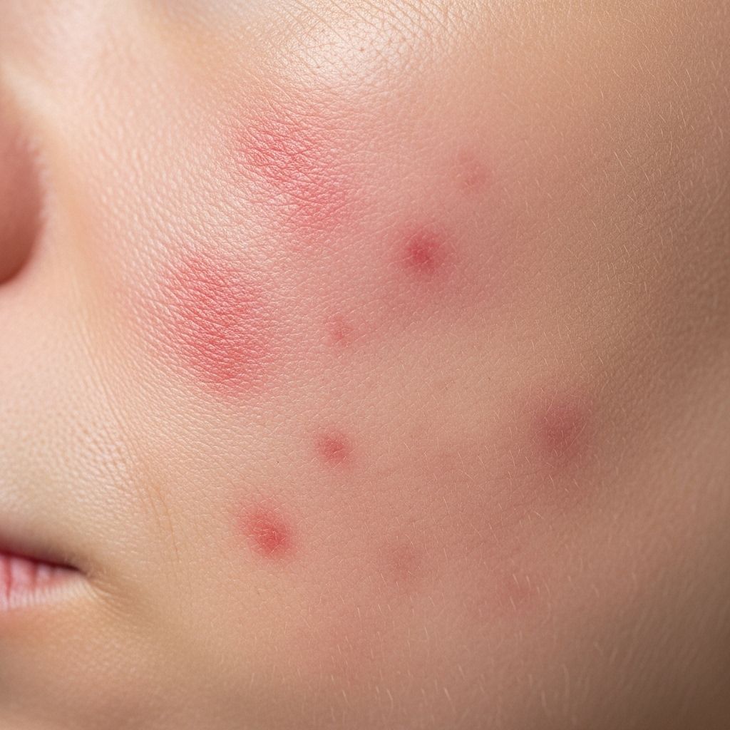
Post-inflammatory erythema (PIE) is one of the most frustrating aftereffects of acne, characterized by persistent pinpoint red marks that linger beneath the skin long after a breakout has healed. PIE not only affects the cosmetic appearance of skin but can also have a psychological effect. In contrast to post-inflammatory hyperpigmentation (PIH), which involves dark brown or black marks due to increased melanin, PIE results from residual inflammation and dilated superficial blood vessels rather than pigment changes.
Table of Contents
- Understanding Post-Inflammatory Erythema
- Causes & Risk Factors of PIE
- PIE vs. PIH
- Timeline & Prognosis
- Topical Treatments for PIE
- Medical & Clinical Treatments
- Prevention & Reduction Strategies
- Frequently Asked Questions (FAQs)
Understanding Post-Inflammatory Erythema
PIE manifests as red or pink marks that linger in areas where acne or other inflammatory skin lesions have healed, typically found on lighter skin types. These marks stem from dilation of capillaries and persistent inflammation—not from pigment changes unlike PIH. PIE can be particularly stubborn, resisting rapid resolution and sometimes persisting for months.
Key Features of PIE:
- Red, pink, or purple spots left behind after inflammatory acne lesions heal.
- Caused by broken or dilated superficial blood vessels and localized inflammation.
- More common in fair or lighter skin tones; darker skin may show more PIH.
- May persist without intervention for 3 to 6 months, but often fades over time in most cases.
Causes & Risk Factors of PIE
PIE is predominantly a consequence of acute inflammation—most often from acne vulgaris. Below are the main risk factors and causes associated with PIE:
- Inflammatory Acne: Severe forms, such as cystic or nodular acne, trigger significant inflammation and disruption of the dermal capillaries.
- Mechanical Trauma: Squeezing, picking, or scratching acne lesions exacerbates vascular injury and increases the risk of PIE.
- Procedures & Irritation: Aggressive skin procedures, chemical peels, or harsh skin products may provoke PIE formation in acne-prone individuals.
- Genetic & Skin Type: Lighter skin types are more likely to exhibit PIE, whereas darker skin may display PIH instead.
PIE vs. PIH: How Is PIE Different?
| Feature | Post-Inflammatory Erythema (PIE) | Post-Inflammatory Hyperpigmentation (PIH) |
|---|---|---|
| Color | Red, pink, or purple | Brown, black, or gray |
| Underlying Cause | Vascular injury, inflammation | Melanin overproduction |
| Common Skin Types Affected | Lighter skin tones | Darker skin tones |
| Treatment Methods | Reduce inflammation, support capillary repair | Reduce melanin, boost cell turnover |
It’s crucial to distinguish between these conditions, as treatments differ: melanin reducers like hydroquinone are ineffective for PIE, which requires anti-inflammatory and vascular-specific therapies.
Timeline & Prognosis: How Long Does PIE Last?
PIE typically evolves as follows:
- Spontaneous resolution may occur in 3–6 months for mild cases as the skin naturally heals and capillaries shrink.
- Persistent PIE, especially after deep cysts or nodules, may linger for 6–12 months or longer.
- Targeted intervention, such as clinical procedures, can greatly accelerate the fading process in most individuals.
Factors influencing prognosis include severity, skin sensitivity, sun exposure, and the use of effective treatments or maintenance routines.
Topical Treatments for PIE
Several topical options may help reduce PIE. Their efficacy varies, and results often depend on the ingredients, formulation, and adherence to routine.
- Topical Hydrocortisone: A mild steroid that reduces inflammation, sometimes prescribed in short bursts. Use only under professional guidance due to risk of skin thinning and side effects.
- Niacinamide: A form of vitamin B3 with anti-inflammatory and antioxidant effects. Regular application can help reduce redness, strengthen the skin barrier, and assist overall healing.
- Vitamin C: An antioxidant shown to lighten pigmentation due to UV damage and may have a mild effect on PIE. It can help brighten and reinforce skin defenses but works best for concurrent PIH.
- Antioxidants (Vitamin E, Green Tea Extract): Neutralize free radicals, support skin repair, and may subtly decrease erythema with consistent use.
- Benzoyl Peroxide & Salicylic Acid: Used primarily to prevent new breakouts, these ingredients fight bacteria and inflammation, helping minimize future PIE formation, though they have limited efficacy for existing marks.
- Retinoids: Including tretinoin and over-the-counter retinol, these increase cell turnover and reduce red marks over weeks or months. They also help prevent recurrence by keeping pores clear.
- Tranexamic Acid, Oxymetazoline, Brimonidine Tartrate: Recent clinical studies show promise for these topical agents in shrinking erythematous lesions by acting directly on vascular processes. However, these may require prescription or physician oversight.
Consistency is essential. Many topical treatments take 8–16 weeks to show results, and irritation should be monitored, particularly with retinoids.
Medical & Clinical Treatments
For persistent PIE that does not respond to topicals, professional procedures can offer substantial results:
- Laser Therapy: Considered the gold standard for resistant PIE. Several modalities are available:
- Pulsed Dye Laser (PDL): Targets blood vessels specifically, improving redness efficiently. Most patients require multiple sessions spaced weeks apart.
- Intense Pulsed Light (IPL): Uses a broad spectrum of light to reduce redness and even skin tone; suitable for lighter skin types.
- Neodymium:yttrium aluminum-garnet (Nd:YAG) Laser: Second in frequency, especially for deeper vessels and more stubborn erythema.
Risks: Pain, transient swelling, risk of pigmentation or scarring, justifying professional oversight.
- Microneedling & Fractional Microneedling Radiofrequency (FMR):
- Tiny needles puncture the skin, inducing collagen production and vascular repair.
- Fractional microneedling radiofrequency (FMR) combines energy-based stimulation, proven to significantly improve PIE within two sessions, as shown in clinical studies. No severe adverse effects observed.
Best performed at a dermatology clinic. Outcomes are both functional and cosmetic, reducing redness and smoothing surface scars.
Prevention & Reduction Strategies
- Treat Active Acne Promptly: Reduces the severity and formation of PIE post-healing.
- Avoid Picking and Squeezing: Trauma to acne lesions exacerbates PIE risk and can lead to scarring.
- Use Sun Protection Every Day: UV light increases skin redness; apply lightweight, non-comedogenic sunscreen suitable for acne-prone skin.
- Adopt Gentle Skin Care Habits: Use non-irritating cleansers; avoid physical scrubs that can worsen inflammation.
- Regular Dermatology Review: Early intervention with a professional prevents stubborn PIE and scarring.
- Maintain Moisture Balance: Hydrated skin heals faster; consider ceramide-rich moisturizers or hyaluronic acid serums.
Frequently Asked Questions (FAQs)
Q: How long does it take for PIE to fade naturally?
A: Most cases of PIE resolve within 3–6 months without intervention, but stubborn marks may last longer depending on severity and skin type.
Q: Will PIE turn into scars if untreated?
A: PIE itself is not scarring, but if underlying inflammation or trauma persists, permanent changes such as atrophic scars can develop. Early management is vital.
Q: Can over-the-counter products help with PIE?
A: Several OTC products containing niacinamide, vitamin C, gentle retinoids, and antioxidants may help reduce PIE over time. Consistency and patience are key.
Q: Is laser therapy safe for all skin types?
A: IPL and PDL are most effective for lighter skin tones. Darker skin types require careful evaluation due to risk of hyperpigmentation or burns. Always consult a board-certified dermatologist.
Q: Are there any home remedies that work?
A: While petroleum jelly, silicone gel sheets, and gentle moisturizers can support healing, evidence for over-the-counter home remedies is limited for PIE; proven results come from clinical-grade topical ingredients and procedures.
Summary Table: Treatments for PIE
| Treatment | Type | Primary Action | Effectiveness | Risks/Considerations |
|---|---|---|---|---|
| Hydrocortisone | Topical | Anti-inflammatory | Moderate | Skin thinning (not for prolonged use) |
| Niacinamide | Topical | Anti-inflammatory, barrier support | Good (for mild PIE) | Low irritation risk |
| Retinoids (Tretinoin, Retinol) | Topical | Increases cell turnover | Good (takes weeks to months) | May cause dryness, irritation |
| Pulsed Dye Laser (PDL) | Clinical | Targets blood vessels | High (gold standard) | Requires multiple sessions, cost |
| Fractional Microneedling Radiofrequency (FMR) | Clinical | Stimulates collagen, vascular repair | High (low risk per studies) | Temporary redness, cost |
| Antioxidants (Vitamin C, E, Green Tea) | Topical | Reduces oxidative stress | Supportive/lightening effect | Low risk |
| Tranexamic acid, Oxymetazoline, Brimonidine tartrate | Topical | Vascular shrinking, anti-inflammatory | Emerging evidence | May require prescription |
Key Sources & Academic Insights
- Laser and light therapies (PDL, IPL, Nd:YAG) have demonstrated the highest efficacy in clinical trials for persistent PIE, often requiring multiple treatments for best results.
- Fractional microneedling radiofrequency (FMR) shows strong promise for rapid PIE reduction with minimal side effects.
- Emerging topicals, such as oxymetazoline and tranexamic acid, act directly on vascular mechanisms to shrink red marks; ongoing studies suggest these options may become mainstream.
- Preventing initial inflammation and trauma, using sunscreen, and supporting skin barrier function are the foundation of reducing PIE formation.
Effective Management of PIE Red Marks: Evidence-Based Care
Post-inflammatory erythema need not be a permanent reminder of past acne. From targeted topical ingredients to cutting-edge clinical treatments, modern dermatology offers numerous options to speed recovery and minimize persistent skin redness. Individuals struggling with PIE should be proactive: adopt evidence-based topical routines, protect their skin from further inflammation, and consult with board-certified dermatologists for advanced procedures when appropriate. With vigilance and science-backed strategies, clear, calm skin is a realistic goal.
References
- https://www.healthline.com/health/acne/post-inflammatory-erythema
- https://slmdskincare.com/blogs/learn/post-inflammatory-erythema-pie-treating-after-acne-red-spots
- https://pubmed.ncbi.nlm.nih.gov/35076997/
- https://www.medicaljournals.se/acta/download/10.2340/00015555-2164/
- https://curology.com/blog/how-to-treat-and-help-prevent-post-inflammatory-erythema/
- https://www.webmd.com/skin-problems-and-treatments/acne/what-to-know-about-post-inflammatory-erythema
- https://naturalimageskincenter.com/post-inflammatory-erythema-vs-post-inflammatory-hyperpigmentation/
- https://pmc.ncbi.nlm.nih.gov/articles/PMC3780804/
- https://www.youtube.com/watch?v=m_8W_mORbmc
Read full bio of medha deb


