Skin Anatomy Basics: Structure, Function, and Roles of the Epidermis, Dermis, and Subcutis
Each tier of your body’s barrier works together to defend, sense and control temperature.

Skin Anatomy Basics: Epidermis, Dermis, Subcutis
The human skin, the body’s largest and most visible organ, serves as the primary interface with the environment. This article provides a detailed overview of the skin’s structure, breaking down its three main layers—the epidermis, dermis, and subcutaneous tissue (subcutis/hypodermis)—and highlights their unique structures, cellular components, and essential physiological functions.
Table of Contents
- Overview of Skin Structure
- Epidermis: The Outer Barrier
- Dermis: The Supportive Connective Layer
- Subcutis (Hypodermis): Fatty Cushion and Energy Reserve
- Functions of the Skin
- Cellular Components of the Skin
- Frequently Asked Questions (FAQs)
Overview of Skin Structure
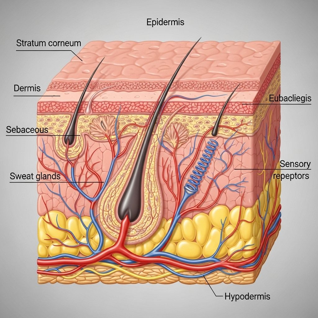
Normal human skin comprises three main layers, each playing distinct roles:
- Epidermis: The superficial, protective barrier.
- Dermis: The underlying supportive connective tissue, vascular and innervated.
- Subcutis (hypodermis): The deepest layer, composed mostly of fat and connective tissue.
| Layer | Main Composition | Key Functions |
|---|---|---|
| Epidermis | Keratinocytes, melanocytes, Langerhans cells, Merkel cells | Barrier, protection, sensation |
| Dermis | Collagen fibers, blood vessels, nerves, glands, fibroblasts | Support, nourishment, elasticity, thermoregulation |
| Subcutis | Fat (adipocytes), connective tissue | Insulation, shock absorption, energy storage |
Epidermis: The Outer Barrier
The epidermis is the skin’s outermost layer. Thin yet resilient, it provides essential protection from environmental assault and contributes to skin tone and waterproofing.
Structural Features of the Epidermis
- Thickness varies widely: thickest on palms (up to 1.5 mm) and soles; thinnest on eyelids and genitalia.
- Entirely avascular—nourished by diffusion from the dermis.
Layers of the Epidermis
From deepest to most superficial, the epidermis consists of:
- Stratum basale (germinativum): Single layer of dividing basal (stem) cells; source of new keratinocytes.
- Stratum spinosum: Several layers of polygonal cells with prominent desmosomes (‘spines’).
- Stratum granulosum: 3–5 cell layers; keratinocytes contain keratohyalin and lamellar granules. These contribute to the formation of the skin’s water barrier and mechanical strength.
- Stratum lucidum (only in thick skin): Thin, translucent layer; found on palms and soles.
- Stratum corneum: Outermost layer; many layers of anucleate, flattened, dead keratinocytes (corneocytes) filled with keratin, providing a tough, water-resistant barrier.
Cell Types in the Epidermis
- Keratinocytes: Main structural cells; produce keratin protein and participate in vitamin D synthesis upon UV exposure.
- Melanocytes: Pigment-producing cells located in the basal layer; synthesize melanin, distributing it to keratinocytes to protect against UV radiation.
- Langerhans cells: Dendritic antigen-presenting cells (immune surveillance), chiefly in the stratum spinosum.
- Merkel cells: Mechanoreceptor cells involved in touch sensation (especially in fingertips).
Function of the Epidermis
- Serves as the body’s primary barrier against microorganisms, chemicals, and physical injury.
- Prevents excess water loss (desiccation) or entry (overhydration).
- Initiates the production of vitamin D from cholesterol precursors via UV light.
- Determines skin color, in conjunction with melanocytes and hemoglobin in underlying vessels.
Dermis: The Supportive Connective Layer
The dermis lies just beneath the epidermis, conferring strength, elasticity, and nourishment to the skin.
Structure and Zones of the Dermis
- Thickness: Varies across the body, from 1–4 mm on average; thickest on the back (almost 1 cm), thinnest on the eyelids.
- Composed mainly of dense connective tissue (primarily types I and III collagen fibers), providing tensile strength.
- Contains elastin fibers (flexibility), ground substance, and an extensive extracellular matrix.
- Houses blood vessels, lymphatics, nerves, and skin appendages (hair follicles, sebaceous/oil glands, sweat glands).
Main Regions of the Dermis
- Papillary Dermis: Superficial, thin layer of loose connective tissue directly under the basement membrane; forms dermal papillae which increase surface area for attachment and nutrient exchange with the epidermis.
- Reticular Dermis: Deeper, thicker region of dense irregular connective tissue; contains larger blood vessels, sweat and sebaceous glands, hair follicles, and fibroblasts.
Functions of the Dermis
- Provides structural support and resilience.
- Nourishes the avascular epidermis via a capillary network.
- Supports cutaneous sensation through a dense innervation of nerve endings and specialized receptors.
- Regulates temperature through vasodilation/vasoconstriction and sweat gland activity.
- Facilitates immune responses; contains immune cells such as dermal dendrocytes and mast cells.
Subcutis (Hypodermis): Fatty Cushion and Energy Reserve
The subcutis (also called the hypodermis or subcutaneous tissue) lies beneath the dermis and forms the deepest layer of skin.
Structure of the Subcutis
- Primarily composed of loose connective tissue and lobules of adipocytes (fat cells).
- Also contains larger blood vessels and nerves that branch upward into the dermis.
- Thickness varies by anatomical site, age, gender, and body composition.
Functions of the Subcutis
- Acts as a thermal insulator, conserving body heat.
- Functions as a shock absorber to protect underlying muscles and bones from trauma.
- Serves as the body’s main energy storage depot.
- An essential site for hormone conversion, such as estrogen production from androgens in adipose tissue.
Functions of the Skin
The skin, in its entirety, is far more than a simple covering—it is a dynamic and multifunctional organ:
- Protection: First physical and immunological barrier against pathogens, mechanical forces, and harmful chemicals.
- Sensation: Houses specialized receptors for heat, cold, pressure, vibration, and pain.
- Thermoregulation: Sweat glands and a rich blood supply regulate heat loss and retention.
- Metabolic functions: Initiates vitamin D production necessary for calcium balance and bone health.
- Excretion: Removes minor waste products (urea, ammonia) through sweat.
- Social and aesthetic roles: Color and condition of skin contribute to individual identity and perception of health.
Cellular Components of the Skin
Each layer of the skin contains a unique combination of cells that reflect its functions:
- Keratinocytes: Predominant epidermal cells responsible for keratin production.
- Melanocytes: Pigment-producing cells in the basal epidermis.
- Langerhans cells: Immunologic sentinels in the epidermis.
- Merkel cells: Sensory cells in the epidermal-dermal junction.
- Fibroblasts: Produce collagen, elastin, and ground substance in the dermis.
- Adipocytes: Fat-storing cells in the hypodermis.
- Immune cells: Mast cells, dendritic cells, and lymphocytes, mostly in the dermis and hypodermis.
Comparative Table: Layers of the Skin
| Layer | Dominant Cell Types | Other Features | Main Functions |
|---|---|---|---|
| Epidermis | Keratinocytes, Melanocytes, Langerhans Cells, Merkel Cells | Multiple strata, Avascular | Barrier, UV defense, Sensation |
| Dermis | Fibroblasts, Immune cells | Dense connective tissue, Glands, Vascular | Support, Nourishment, Elasticity, Sensation |
| Subcutis | Adipocytes, Few fibroblasts | Loose connective tissue, Vascular, Nerve fibers | Insulation, Energy store, Shock absorber |
Frequently Asked Questions (FAQs)
Q: What are the main differences between the epidermis and dermis?
The epidermis is a thin, outermost, avascular layer composed mainly of keratinocytes, providing a waterproof barrier and physical protection. The dermis is thicker, vascular, and made up of connective tissue, housing essential structures such as blood vessels, nerves, glands, and hair follicles. The dermis provides structural support and elasticity.
Q: Is the hypodermis the same as subcutaneous tissue?
Yes. The terms hypodermis and subcutaneous tissue (subcutis) are used interchangeably to describe the deepest layer of skin, mainly consisting of adipose tissue and loose connective tissue.
Q: How does skin protect the body from diseases?
The skin serves as the first line of defense, forming a physical barrier to bacteria, viruses, fungi, and toxins, and hosting immune cells (such as Langerhans cells and mast cells) that detect and respond to invading pathogens.
Q: How is vitamin D produced in the skin?
Vitamin D synthesis begins when the skin’s keratinocytes absorb ultraviolet B (UVB) radiation, converting 7-dehydrocholesterol to pre-vitamin D3, which is further processed in the liver and kidneys to the active hormone.
Q: Why does the skin have different thicknesses?
Skin thickness varies to accommodate function and exposure: areas exposed to friction (palms, soles) have thick skin to withstand mechanical forces, while the eyelids and genitalia feature thinner skin for sensitivity and flexibility.
Q: What causes differences in skin color?
Skin color is determined primarily by the amount, type, and distribution of melanin produced by melanocytes, influenced by genetics and UV exposure. Vascularization and carotene pigment also contribute to tone and pigmentation.
Summary
The human skin is a complex, multitiered organ crucial for health and well-being. Its three main layers—the epidermis, dermis, and subcutis—work as an integrated system to protect the body, mediate sensation, regulate temperature, support metabolism, and express identity. A fundamental understanding of skin anatomy is essential for recognizing how this vital organ guards us against the external world, maintains internal stability, and reflects overall health.
References
- https://www.utmb.edu/pedi_ed/CoreV2/Dermatology/page_03.htm
- https://www.ncbi.nlm.nih.gov/books/NBK441980/
- https://www.ncbi.nlm.nih.gov/books/NBK470464/
- https://journals.cambridgemedia.com.au/wpr/volume-32-number-1/anatomy-physiology-and-function-all-skin-layers-and-impact-ageing-skin
- https://my.clevelandclinic.org/health/body/10978-skin
- https://www.stanfordchildrens.org/en/topic/default%3Fid=anatomy-of-the-skin-85-P01336
- https://my.clevelandclinic.org/health/body/21902-hypodermis-subcutaneous-tissue
- https://teachmeanatomy.info/the-basics/ultrastructure/skin/
Read full bio of medha deb


