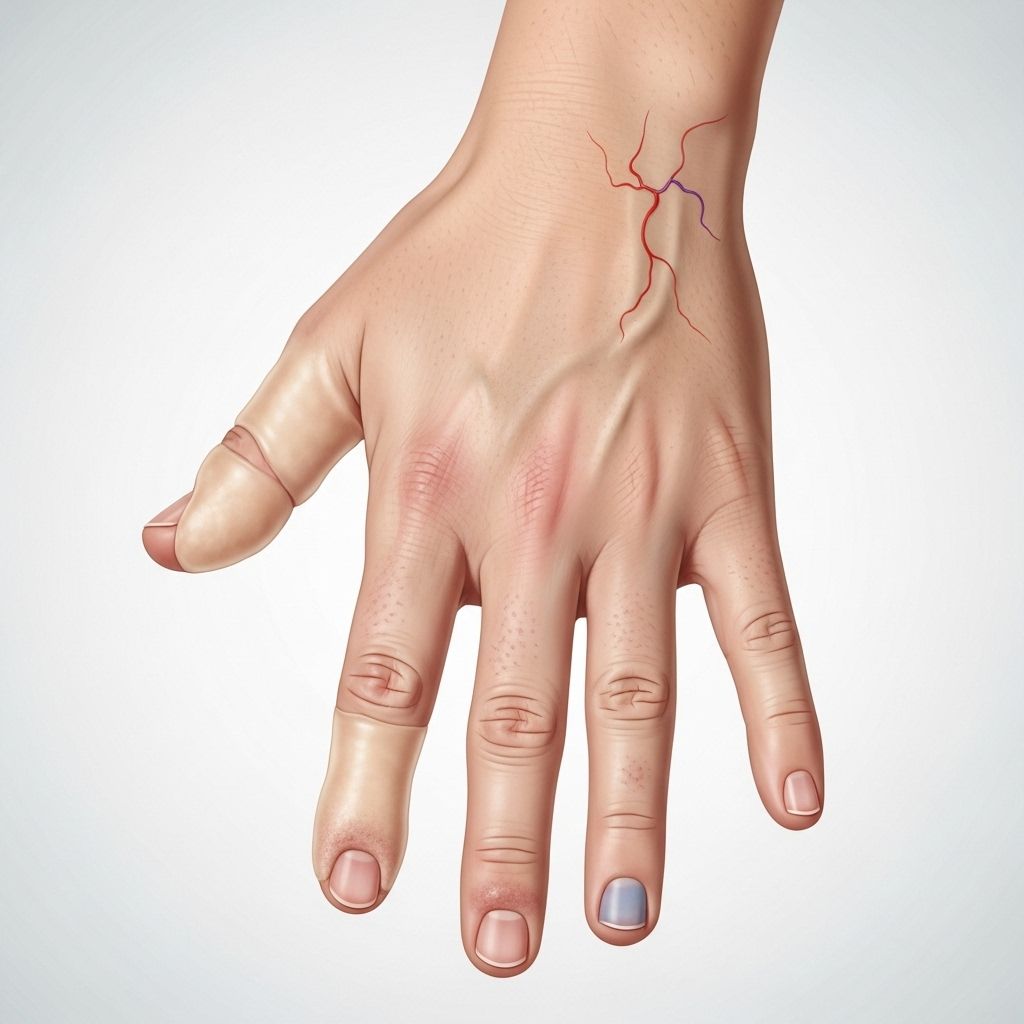
Recognizing and Managing Scleroderma Skin Changes: Detailed Strategies, Signs, and Patient Care
Spotting subtle signs early empowers better treatment and long-term comfort.

Recognizing and Managing Scleroderma Skin Changes
Scleroderma is a complex autoimmune condition that most often affects the skin but can impact other organs as well. The term scleroderma literally means “hard skin” and refers to the key symptom of skin thickening from excess collagen production. Recognizing early skin changes and implementing targeted management strategies is vital for improving outcomes and quality of life for those affected by this chronic disease.
Table of Contents
- What is Scleroderma?
- Types of Scleroderma and Associated Skin Changes
- Recognizing Scleroderma Skin Changes
- Diagnosis: Approaches and Tests
- Managing Scleroderma Skin Changes
- Potential Complications and Prevention
- Living with Scleroderma: Daily Life & Support
- Frequently Asked Questions (FAQs)
What is Scleroderma?
Scleroderma is a chronic autoimmune disease characterized primarily by inflammation and abnormal growth of connective tissue, resulting in skin thickening (fibrosis) and sometimes organ involvement. In scleroderma, the body’s immune system mistakenly attacks its own healthy tissues, leading to excess collagen production and tissue hardening.
- Most frequently affects the skin, but may involve blood vessels and internal organs.
- More common in adult women, but localized forms often affect children.
- Its severity and progression vary widely among individuals.
Types of Scleroderma and Associated Skin Changes
Scleroderma is divided into localized and systemic forms, each with subtypes and distinct patterns of skin involvement.
Localized Scleroderma
- Morphea: Causes patches of thick, waxy, discolored skin on the chest, back, stomach, arms, or legs. These may enlarge, change shape, or resolve over time.
- Linear scleroderma: Manifests as streaks of hardened, shiny skin, typically on the limbs or forehead (“en coup de sabre”), and can restrict joint movement, impact limb growth in children, and affect deeper tissues.
Systemic Scleroderma (Systemic Sclerosis)
- Limited cutaneous scleroderma (CREST syndrome): Most often affects fingers, hands, face, forearms, and legs below the knees. May cause vessel and esophageal issues, and develop slowly.
- Diffuse cutaneous scleroderma: Skin thickening begins in the hands or feet and rapidly spreads to larger body areas, including trunk and face, with rapid progression and risk of organ involvement.
- Sine sclerosis: Internal organ damage occurs without noticeable skin thickening, an uncommon variant.
| Type | Most Affected Areas | Skin Changes |
|---|---|---|
| Morphea (Localized) | Chest, back, legs, arms | Patches, thickening, waxy discoloration |
| Linear (Localized) | Limbs, face | Streaks, shiny/hardened skin, possible joint restriction |
| Limited (Systemic) | Fingers, hands, face, forearms, lower legs | Gradual thickening, dull/shiny appearance, telangiectasia |
| Diffuse (Systemic) | Whole body (above/below elbows/knees, trunk, face) | Rapid progression, widespread skin hardening |
| Sine Sclerosis (Systemic) | Internal organs only | No obvious skin changes |
Recognizing Scleroderma Skin Changes
Early detection and recognition of skin changes in scleroderma are essential, as interventions may reduce progression and complications. Clinical evaluation relies upon both visual inspection and symptom reports.
Common Skin Symptoms
- Hardening and tightening of skin: Skin feels firm, immobile, and may appear shiny due to tightness.
- Swelling and puffiness: Early signs, especially in fingers and toes—sometimes preceding skin hardening.
- Itching and dryness: May accompany swelling, particularly in initial phases.
- Change in skin color: Affected skin may be darker (hyperpigmentation) or lighter (hypopigmentation), or have a waxy, yellowish tinge.
- Loss of hair: Hairless patches can develop on thickened areas.
- Ulcerations and pits: Especially on fingertips, elbows, or ankles, due to tissue loss and poor circulation.
- Telangiectasia: Small, dilated red blood vessels often visible on hands and face.
- Mask-like facial appearance: Severe skin tightening can reduce facial expressions and mouth opening.
- Calcinosis cutis: Hard, white or yellow lumps (calcium deposits) under the skin, typically in advanced disease.
Symptoms Beyond the Skin
- Restricted joint mobility (contractures)
- Muscle weakness and limb deformities (particularly in children)
- Raynaud’s phenomenon (pain, color changes in fingertips/toes due to poor blood flow)
Diagnosis: Approaches and Tests
The clinical suspicion of scleroderma skin changes prompts further assessments for accurate diagnosis and monitoring. Diagnosis typically combines history, physical exam, and supportive laboratory or imaging studies.
- Skin examination: Detects location, pattern, and severity of hardening or color changes.
- Medical history: Focuses on symptom duration, presence of Raynaud’s phenomenon, systemic complaints, and family history.
- Skin biopsy: May confirm scleroderma and assess disease type.
- Blood tests: Evaluate for specific autoantibodies (e.g., anti-centromere, anti-Scl-70), organ function, and inflammation.
- Imaging: X-rays or ultrasound for calcinosis, assessment of joint changes, and organ involvement.
- Nailfold capillaroscopy: Visualizes microvascular changes under fingernails, assisting with systemic diagnosis.
Timely recognition and diagnostic clarity underpin effective management strategies.
Managing Scleroderma Skin Changes
While no cure exists for scleroderma, management focuses on controlling symptoms, preventing progression, minimizing complications, and supporting overall skin and joint health. Therapy is individualized based on symptoms, disease severity, and extent of organ involvement.
Medical Treatments
- Topical treatments: Emollients, corticosteroid creams, and calcineurin inhibitors to reduce inflammation and maintain skin moisture.
- Phototherapy: Ultraviolet light (UVA1 phototherapy) used for localized skin involvement (especially morphea).
- Systemic immunosuppressants: Drugs like methotrexate, mycophenolate mofetil, or cyclophosphamide (for more aggressive or widespread skin involvement).
- Calcium channel blockers: For Raynaud’s phenomenon, improving blood flow and reducing episode frequency.
- Medications for ulcerations and calcinosis: Specific interventions to heal persistent ulcers or manage calcium deposits.
Physical and Supportive Therapies
- Physical therapy: Maintains joint mobility, prevents contractures, and improves limb function.
- Occupational therapy: Facilitates adaptive strategies for daily activities given skin and hand changes.
- Protective measures: Routine skin care, gentle cleansing, and protection from trauma and extreme temperatures.
Self-care Strategies
- Keep skin well-hydrated with rich, unscented moisturizers.
- Avoid harsh soaps or hot water, which can worsen dryness.
- Use gloves and protective gear during household chores, gardening, or cold weather.
- Regular gentle stretching to maintain flexibility.
- Inspect skin daily for ulcerations, infection, or color changes.
Potential Complications and Prevention
Effective management seeks both to preserve skin integrity and minimize complications commonly associated with progressive scleroderma.
Possible Skin Complications
- Skin ulcerations: Slow-healing wounds, mostly on fingertips.
- Calcinosis cutis: Painful calcium deposits may ulcerate or become infected.
- Pitting and scarring: Irregularities and tissue loss may occur, especially in longstanding cases.
- Telangiectasia bleeding: Surface blood vessels may rupture, causing minor bleeding.
- Skin infections: Compromised skin is more prone to infections.
- Contractures: Joint immobility due to thickened skin and connective tissue around joints.
Prevention and Monitoring
- Prompt management of ulcers, ideally with the support of a wound care team.
- Regular dermatology and rheumatology follow-up.
- Education on signs of infection for early intervention.
- Minimize trauma with protective behaviors.
- Monitor and address systemic complications (pulmonary, renal, cardiac) in systemic forms.
Living with Scleroderma: Daily Life & Support
Chronic skin changes can significantly affect quality of life—both physically and emotionally. Comprehensive care, education, and psychosocial support enhance well-being and resilience.
Psychosocial Support
- Counseling services to address self-esteem, depression, or body image concerns.
- Local and online support groups for information sharing and community.
- Education on adaptation strategies for activities of daily living.
Patient Education
- Understanding the disease process and realistic goals for therapy.
- Awareness of potential complications and the importance of self-monitoring.
- Empowering patients to communicate changes and challenges with healthcare providers.
Integrative Approaches
- Relaxation exercises, gentle yoga, or mindfulness to reduce stress (which may trigger flares).
- Diet modifications if digestive symptoms are present.
- Sun protection to prevent additional skin damage and pigment changes.
Frequently Asked Questions (FAQs)
Q: What are the first signs of scleroderma skin involvement?
Early signs commonly include puffiness, swelling (especially of fingers and toes), followed by hardening, tightening, and shiny appearance of skin, often accompanied by color change or itching.
Q: How is scleroderma skin involvement diagnosed?
Diagnosis involves physical examination, symptom history, skin biopsy (when needed), blood tests for autoantibodies, and occasionally imaging or capillary studies.
Q: Can scleroderma skin changes improve or reverse?
Some localized forms (such as morphea) may stabilize or improve; systemic sclerosis rarely reverses but progression can be slowed with treatment. Timely and consistent management is critical for best outcomes.
Q: What daily care strategies help manage scleroderma skin changes?
- Hydrate skin regularly with rich moisturizers.
- Avoid trauma and cold exposure to hands and feet.
- Regular gentle stretching exercises to maintain joint mobility.
- Monitor for wounds or infections; seek prompt medical attention for ulcers.
Q: Is scleroderma contagious or inherited?
No, scleroderma is not contagious nor directly inherited, but genetic and environmental factors may increase risk.
Summary and Key Points
- Scleroderma is a chronic autoimmune disease characterized by skin thickening and hardening, often accompanied by color and textural changes.
- Two main types—localized and systemic—cause distinct patterns of symptoms and risks.
- Early recognition of skin changes allows for timely intervention, minimizing complications and improving quality of life.
- No cure exists, but skin changes are manageable with topical, systemic, physical, and supportive therapies tailored to each individual’s needs.
- Comprehensive care—including psychosocial support, patient education, and lifestyle adaptation—plays a vital role in successful management and community integration.
References
- https://www.yalemedicine.org/conditions/scleroderma
- https://www.niams.nih.gov/health-topics/scleroderma
- https://www.mayoclinic.org/diseases-conditions/scleroderma/symptoms-causes/syc-20351952
- https://www.pennmedicine.org/conditions/scleroderma
- https://my.clevelandclinic.org/health/diseases/scleroderma
- https://scleroderma.org/symptoms-of-scleroderma/
- https://scleroderma.org/types-of-scleroderma/
- https://www.sruk.co.uk/about-scleroderma/signs-symptoms-of-scleroderma/effects-of-scleroderma-on-the-body/skin-in-systemic-sclerosis/
Read full bio of Sneha Tete


