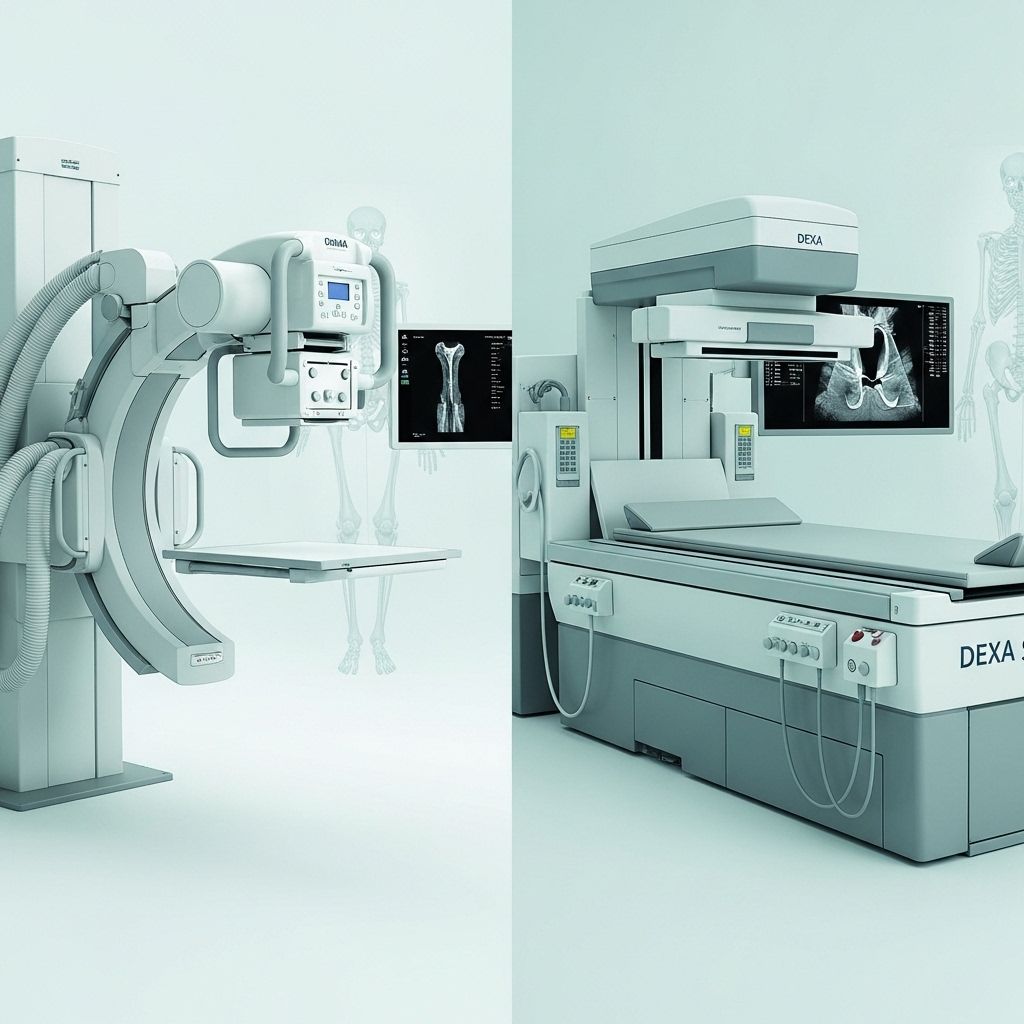
Comprehensive Comparison of Radiography and Dexa Scan for Bone Density Assessment: Methods, Accuracy, and Clinical Utility
Choosing the most precise imaging method improves diagnosis and informs treatment plans.

Radiography vs. Dexa Scan Bone Density Comparison
With the increasing emphasis on early detection and management of osteoporosis and other bone health conditions, medical practitioners rely heavily on imaging for bone assessment. Two commonly used modalities are radiography (standard x-ray imaging) and DEXA scan (dual-energy x-ray absorptiometry). This article offers a detailed comparison of these methodologies, outlining their principles, accuracy, clinical relevance, benefits, limitations, and practical considerations.
Table of Contents
- Overview of Bone Imaging Techniques
- Technical Principles
- Clinical Applications
- Accuracy and Sensitivity
- Procedural Experience
- Benefits and Risks
- Comparison Table: Radiography vs. DEXA Scan
- Recent Advances and Research Insights
- Limitations and Considerations
- Frequently Asked Questions (FAQs)
Overview of Bone Imaging Techniques
Bone imaging plays a pivotal role in diagnosing fractures, bone deformities, metabolic bone disorders, and osteoporosis. While both radiography and DEXA utilize x-ray technology, they differ markedly in their diagnostic capabilities and clinical focus.
- Radiography is the traditional method for visualizing bone structure, shape, alignment, and acute pathology.
- DEXA scan specifically measures bone mineral density (BMD), providing quantitative data for osteoporosis assessment and fracture risk evaluation.
Technical Principles
Radiography
Radiography uses a single energy x-ray beam to capture images of teeth, bones, and tissues. The images produced are two-dimensional representations, which show bone anatomy but do not quantitatively measure bone density.
- The beam passes through the body, and denser materials like bone absorb more x-rays, appearing whiter on film.
- Used to detect fractures, joint abnormalities, tumors, and infections.
- Cannot reliably disclose minor variations in bone density, particularly in early stages of osteoporosis.
DEXA Scan (Dual-Energy X-ray Absorptiometry)
DEXA employs two x-ray beams at different energy levels. By measuring the differential absorption, it can determine the density of bones with high precision.
- The two energy levels enable subtraction of soft tissue absorption, leaving an accurate measurement of bone mineral content.
- DEXA is considered the gold standard for quantifying BMD and tracking changes over time.
- Most commonly used for lumbar spine and hip assessment—critical areas for detecting osteoporosis.
Clinical Applications
- Radiography
- Fracture detection (acute and chronic)
- Bone infection and tumor identification
- Skeletal deformity assessment
- Bony changes in arthritis
- DEXA Scan
- Diagnosis of osteoporosis and osteopenia
- Estimation of fracture risk
- Monitoring response to osteoporosis treatment
- Screening populations at risk (post-menopausal women, elderly, steroid users)
- Peripheral DEXA for forearm, hand, or foot assessment in some cases
Accuracy and Sensitivity
The accuracy and clinical value of an imaging modality determine its effectiveness in guiding care. DEXA vastly outperforms radiography in sensitivity to bone mineral content, particularly in early disease.
- Radiography limits: Plain x-rays only detect bone loss once it exceeds 30–40%. This means significant osteoporosis can exist before radiographs show noticeable changes.
- DEXA strengths: DEXA scans can detect subtle reductions in bone mineral density, enabling early diagnosis and intervention—crucial for preventing fractures in at-risk populations.
For pediatric populations, research has shown that DEXA (particularly with iDXA systems) can accurately assess bone age and even vertebral morphometry, sometimes replacing conventional radiography and lowering radiation exposure.
Procedural Experience
Radiography
Standard x-ray exams are quick (usually under 10 minutes), require little preparation, and involve positioning the body part of interest near the x-ray source. The patient must remain still to get a clear image.
- Non-invasive and generally painless
- Minimal preparation: remove jewelry, wear a simple gown if needed
DEXA Scan
DEXA scans are also non-invasive and quick, typically completed within 10–30 minutes depending on the anatomic site and equipment.
- Patient lies on padded table, remaining still while a scanner passes over spine or hip.
- No food, drink, or calcium supplements 24 hours before test.
- Peripheral DEXA scans (forearm, finger, ankle) may be used in some settings.
Patients may undergo a short questionnaire for fracture risk modeling; results are interpreted by radiologists and shared with referring doctors.
Benefits and Risks
Radiography
- Benefits: Rapid diagnosis of acute pathology; widely available and inexpensive.
- Risks: Moderate radiation exposure; cannot diagnose mild osteopenia or early osteoporosis.
DEXA Scan
- Benefits: Minimal radiation (much less than standard x-rays); high sensitivity and specificity for BMD; direct fracture risk prediction.
- Risks: Rare allergic reactions to contrast (only if used in adjunct imaging, not standard DEXA); slightly higher cost.
Comparison Table: Radiography vs. DEXA Scan
| Feature | Radiography (X-ray) | DEXA Scan |
|---|---|---|
| Primary Purpose | Visualize bone structure, detect fractures, general pathology | Quantify bone mineral density, diagnose osteoporosis |
| Technology | Single-energy x-ray beam | Dual-energy x-ray beams |
| Sensitivity to Bone Loss | Low: >30–40% loss needed for detection | High: Detects minor loss and early disease |
| Radiation Dose | Moderate | Low |
| Time Required | 5–15 minutes | 10–30 minutes |
| Diagnostic Value | Bones, fractures, tumors, infection, deformity | Bone density, osteoporosis, fracture risk |
| Limitations | Not sensitive to early bone loss | Does not confirm acute fractures |
Recent Advances and Research Insights
Several research studies have compared the utility of DEXA and conventional radiographs for various indications. Pediatric data indicate that when assessing conditions like osteogenesis imperfecta or secondary osteoporosis, iDXA scans provide comparable results for morphometric assessment and bone age, sometimes allowing replacement of traditional x-ray and reducing radiation exposure.
Modern DEXA technology incorporates vertebral fracture assessment (VFA), which screens for fractures with enhanced sensitivity and minimal additional exposure. Integration of computer algorithms with DEXA enables fracture risk predictions for the next decade using input such as age, sex, prior fractures, medication use, and BMD scores.
Limitations and Considerations
- Radiography remains essential for evaluating acute trauma, bone tumors, or infections, where rapid structural imaging is crucial.
- DEXA has limited spatial resolution, making it unsuitable for diagnosing fractures.
- Not all clinics have access to advanced DEXA scanners; accessibility and cost may be limiting factors in some regions.
- Pregnant patients should avoid both types of scans unless medically necessary, due to ionizing radiation.
- Recent contrast studies or barium meals may interfere with DEXA results.
Frequently Asked Questions (FAQs)
Q: What does a DEXA scan measure that a standard x-ray does not?
A: DEXA provides a quantitative assessment of bone mineral density, detecting early osteoporosis and calculating fracture risk, while standard x-ray only reveals structural bone abnormalities after substantial bone loss.
Q: Is the radiation dose from DEXA higher than that from radiography?
A: No. The radiation dose in DEXA scans is considerably lower than conventional x-rays, often described as similar to daily natural background exposure.
Q: Can DEXA diagnose fractures like an x-ray?
A: No. DEXA is not designed for fracture detection. It evaluates density and risk, whereas x-ray confirms fracture presence and location quickly.
Q: Who should have a DEXA scan?
A: DEXA is recommended for those at high risk for osteoporosis—postmenopausal women, older men, individuals on long-term steroids, or those with history of low-trauma fracture.
Q: How should patients prepare for a DEXA scan or x-ray?
A: For DEXA, avoid calcium supplements and contrast studies for 24 hours; wear comfortable clothing free of metal. For x-ray, remove jewelry and inform the technologist of pregnancy.
Conclusion
Both radiography and DEXA scans are indispensable tools in modern bone health management. While radiography excels in acute structural imaging, DEXA stands alone in providing precise bone density measurement and risk stratification for fragility fractures. In practice, the two techniques are complementary—each with unique strengths, limitations, and applications, contributing synergistically to patient care and prevention strategies for osteoporosis and bone-related pathologies.
References
- https://www.radiologyinfo.org/en/info/dexa
- https://www.patientcarenowurgentcare.com/2024/03/08/bone-density-scans-vs-x-rays-understanding-the-differences/
- https://pubmed.ncbi.nlm.nih.gov/26059565/
- https://mediccloud.com.au/dexa-systems-versus-x-ray-technology/
- https://pmc.ncbi.nlm.nih.gov/articles/PMC9674387/
- https://bergmanross.co.za/bone-density-scan-vs-x-rays-main-cost-differences/
Read full bio of medha deb


