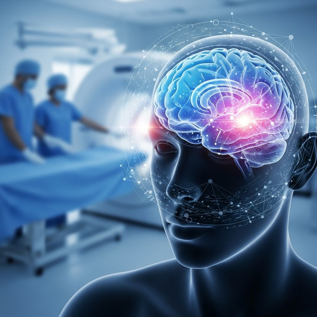
Protocols for Atypical Stroke Symptoms: Detection, Diagnosis, and Emergency Response Strategies
Broadened triage alongside imaging reveals subtle signs overlooked in emergency care.

Atypical stroke symptoms challenge both prehospital and emergency care teams due to their diverse and sometimes subtle presentations. Failure to promptly recognize and respond to atypical strokes—particularly posterior circulation and ‘stroke mimic’ scenarios—can result in delayed treatment, increased morbidity, and preventable mortality. This article presents a detailed review of current protocols, diagnostic pathways, and multidisciplinary approaches to optimize the management of atypical strokes in urgent care settings.
Table of Contents
- Introduction
- Understanding Atypical Stroke Symptoms
- Clinical Challenges in Atypical Stroke Presentations
- Initial Assessment and Triage Protocols
- Diagnostic Strategies and Imaging Protocols
- Stroke Mimics: Differentiation and Clinical Clues
- Emergency Response and Interdisciplinary Management
- Quality Improvement, Education, and Future Directions
- Frequently Asked Questions (FAQs)
Introduction
Stroke remains a leading cause of fatality and long-term disability worldwide. Standard protocols for stroke identification typically rely on classic neurological presentations—sudden facial droop, arm weakness, and speech disturbance. However, atypical stroke symptoms—such as isolated vertigo, visual changes, or nonspecific sensory deficits—can complicate clinical recognition, especially in posterior circulation strokes or presentations involving stroke mimics.
Rapid identification and appropriate triage are critical, as time-sensitive therapies such as thrombolysis and thrombectomy offer maximal benefits when initiated soon after symptom onset. In this context, structured protocols that account for atypical features are essential for improving outcomes.
Understanding Atypical Stroke Symptoms
Atypical strokes often involve non-classical symptoms, making them harder to diagnose than typical ischemic or hemorrhagic strokes. These presentations are more prevalent in certain stroke types—most notably, those affecting the posterior circulation (brainstem, cerebellum, occipital lobes) and in specific populations (elderly, women, or those with prior neurological conditions).
Main Atypical Symptom Categories
- Vertigo and Ataxia: Unsteady gait, dizziness, or sudden inability to walk, sometimes mistaken for vestibular disorders.
- Nausea/Vomiting: Frequently seen with brainstem involvement, sometimes the primary complaint in posterior circulation strokes.
- Isolated Visual Changes: Vision loss, double vision, or field deficits can be sole manifestations, especially in occipital or brainstem strokes.
- Headache and Altered Sensorium: Severe headache without neurological deficit, or confusion and agitation, often confound diagnosis.
- Nonfocal or Fluctuating Deficits: Sensory changes, weakness not fitting a single vascular territory, or rapidly changing symptoms.
In contrast to the typical F.A.S.T. (Face, Arm, Speech, Time) algorithm, atypical symptoms often escape rapid detection, necessitating broadened awareness and expanded triage tools.
Clinical Challenges in Atypical Stroke Presentations
Atypical stroke symptoms introduce significant diagnostic complexity due to their overlap with more benign or nonvascular neurological disorders. Clinical research highlights several challenges:
- Misclassification at Triage: Atypical presentations (e.g., ataxia, dizziness) can be attributed to inner ear disease, migraine, hypoglycemia, or even psychiatric syndromes, leading to diagnostic delays.
- Knowledge Gaps: Emergency physicians and first responders may be less familiar with posterior circulation symptoms, reducing sensitivity in initial stroke screens.
- Systemic Barriers: Protocols often prioritize classic stroke signs, resulting in lower triage scores or delayed imaging for atypically presenting patients.
- Subtlety and Fluctuation: Symptoms that are transient or wax and wane increase the risk of misdiagnosis.
The clinical imperative, therefore, is to incorporate protocol modifications and targeted training to improve recognition.
Initial Assessment and Triage Protocols
Effective management begins at first medical contact, often in the emergency department (ED). Standardized protocols should be broadened to encompass atypical features.
Essential Steps in Triage and Initial Assessment
- History and Onset: Detailed recording of time of symptom onset or “last known well” status is critical for eligibility for acute interventions.
- Immediate Vitals and Glucose Testing: Assess for hypoglycemia or hemodynamic instability (common stroke mimics).
- Expanded Neurological Exam: Go beyond hemiparesis and speech: assess for nystagmus, ataxia, visual field deficits, or cranial nerve abnormalities.
- Screen for Atypical Features: Inquire about dizziness, gait alteration, isolated vision changes, severe headache, nausea/vomiting, or confusion.
- Use Enhanced Triage Tools: Emerging screening checklists like FAST HAND-ED include atypical symptoms (e.g. ataxia, nausea/vomiting, double vision) and can reduce delays.
Ultimately, ED staff must maintain a high index of suspicion, especially when high-risk profiles or sudden-onset neurological deficits do not conform to classic stroke patterns.
Diagnostic Strategies and Imaging Protocols
Early and accurate diagnosis of stroke, especially with atypical symptoms, is crucial to guide therapeutic interventions. The diagnostic process leverages both clinical examination and neuroimaging.
Key Diagnostic Modalities
- Non-Contrast CT Scan: First-line imaging to rule out hemorrhage or large infarct. May be normal in early ischemic stroke or in stroke mimics.
- CT Angiography (CTA): Assesses for large vessel occlusion, vessel dissection, or vascular malformations.
- CT Perfusion (CTP): Useful in extending time windows for intervention based on penumbra-core mismatch.
- Diffusion-Weighted MRI (DWI-MRI): Most sensitive for detecting early ischemic changes, crucial in subtle or fluctuating symptoms, but less readily available.
- Magnetic Resonance Angiography (MRA): Visualizes arterial flow and occlusions, especially valuable for posterior circulation evaluation.
- Carotid Ultrasound and Echocardiography: Evaluate embolic sources if suggestive findings arise.
- Blood Work: Coagulation profile, glucose, electrolytes, infectious/inflammatory markers to rule out mimics.
Emerging expedited protocols now recommend parallel processing (simultaneous clinical exam, labs, and imaging) to minimize delays in diagnosis when atypical symptoms suggest a possible stroke.
Table: Comparison of Imaging Modalities in Acute Stroke
| Imaging Modality | Best For | Advantages | Limitations |
|---|---|---|---|
| Non-Contrast CT | Initial differentiation of hemorrhage vs. ischemic stroke | Widespread availability, rapid | Often negative in early ischemic changes |
| CT-Angiography | Detects vessel occlusion, dissection | Visualizes vasculature quickly | Contrast risks; limited detail in smaller vessels |
| DWI-MRI | Early ischemic changes, subtle deficits | Highest sensitivity | Limited urgent access, contraindications (pacemaker, metal) |
| Carotid Ultrasound | Carotid artery disease | Non-invasive, rapid at bedside | Examiner-dependent, does not evaluate intracranial arteries |
Stroke Mimics: Differentiation and Clinical Clues
Stroke mimics—conditions presenting with similar acute neurological findings but not caused by cerebrovascular pathology—constitute up to 20–30% of stroke code activations. Differentiating these is critical, as mistreatment (e.g., unnecessary thrombolysis) carries unnecessary risk.
Common Stroke Mimics
- Migraine with Aura: Gradual onset, visual phenomena, normal imaging.
- Seizures/Post-ictal States: Transient deficits (e.g., Todd’s paralysis), atypical EEG findings.
- Hypoglycemia: Rapid improvement with glucose correction.
- Functional/Conversion Disorders: Inconsistent exam findings, awareness of surroundings, no imaging abnormality.
- Brain Tumors or Infections: Progressive, less abrupt onset; revealed on imaging over time.
Clinical Clues Distinguishing Mimics from True Stroke
- Gradual onset or fluctuating symptoms
- Symptoms not anatomically distinct or vascular-territory confined
- Improvement with basic ED interventions (e.g., glucose)
- Normal early imaging with progressive changes
Systematic clinical and radiological assessment, with careful attention to red-flag features (sudden onset, maximal deficit at onset, risk factors), is key to distinguishing mimics.
Emergency Response and Interdisciplinary Management
Once suspicion is confirmed, protocols for acute management should be rapidly enacted regardless of symptom typicality. Multidisciplinary teamwork ensures optimized intervention.
Acute Management Protocols
- Code Stroke Activation: Immediate notification of stroke team and radiology.
- Targeted Laboratory Panels: Rapid blood glucose, coagulation panel, and infection/inflammation markers.
- Imaging: Non-contrast CT within minutes; MRI as adjunct where available.
- Neuroconsultation: Early neurologist involvement for complex/atypical cases.
- Thrombolysis/Thrombectomy Timing: Strict assessment for eligibility based on time from onset or advanced imaging findings.
- Address Underlying Etiologies: Manage hypertension, arrhythmias, glucose, and emergent comorbidities.
If stroke mimics are confirmed, further targeted management (e.g., antiepileptics, migraine therapy, or psychiatric referral) is required. Ongoing communication between ED, neurology, radiology, and nursing is mandatory for seamless care transitions.
Quality Improvement, Education, and Future Directions
Quality improvement initiatives and ongoing professional education are vital components of an effective atypical stroke protocol. Recent strategies include:
- Simulation-Based Training: Enhances clinician recognition of non-classic deficits.
- Protocol Audit and Feedback: Regular review of stroke codes for missed or delayed atypical cases.
- Development of Specialized Triage Tools: Wider adoption of expanded algorithms (such as FAST HAND-ED) to capture broader symptom sets.
- Patient and Public Education: Outreach to inform at-risk populations about atypical symptoms, aiming to reduce prehospital delays.
Further research is needed to validate new screening tools and optimize workflow integration for atypical stroke protocols.
Frequently Asked Questions (FAQs)
What are the most common atypical symptoms of acute stroke?
The most frequent atypical symptoms include sudden vertigo, severe dizziness, unsteady gait (ataxia), nausea/vomiting, isolated visual disturbances, and confusion or sudden loss of consciousness. These symptoms frequently signal posterior circulation involvement.
How can atypical stroke symptoms be distinguished from stroke mimics?
Clinical clues such as abrupt onset, maximal symptoms at onset, and anatomical distribution aligned with vascular territories favor true stroke. Fluctuating or gradual symptoms, inconsistency on examination, or rapid deficit resolution often indicate mimics like migraine, seizures, or functional disorders.
Which diagnostic tests are prioritized when atypical symptoms are present?
Non-contrast CT is nearly always first, followed by CT angiography or MRI (if available and warranted). Where suspicion remains, carotid ultrasound, laboratory screening (glucose, coagulation), and echocardiography may assist diagnosis.
What is the role of expanded triage tools like FAST HAND-ED?
Such tools incorporate additional symptoms typical of posterior strokes—ataxia, double vision, nausea/vomiting—and improve early detection and prioritization of care for patients less likely to present with classic symptoms.
Is it possible for imaging to appear normal in patients with atypical stroke symptoms?
Yes, especially in the immediate hours after symptom onset, early CT or MRI scans may show no acute changes. Repeat imaging and careful clinical follow-up are crucial in such cases.
References
- Stroke mimics: incidence, aetiology, clinical features and treatment. PMC.
- Mayo Clinic: Stroke – Diagnosis and treatment.
- Decreasing Delayed Recognition, Diagnosis, & Treatment of Posterior Circulation Strokes. Neurology.org.
- Acute Stroke Diagnosis. AAFP.
References
- https://pmc.ncbi.nlm.nih.gov/articles/PMC7939567/
- https://www.mayoclinic.org/diseases-conditions/stroke/diagnosis-treatment/drc-20350119
- https://www.neurology.org/doi/10.1212/WNL.98.18_supplement.3659
- https://www.aafp.org/pubs/afp/issues/2022/0600/p616.html
- https://pmc.ncbi.nlm.nih.gov/articles/PMC9796438/
- https://www.strokebestpractices.ca/recommendations/secondary-prevention-of-stroke/triage-and-initial-diagnostic-evaluation-of-transient-ischemic-attack-and-non-disabling-stroke
Read full bio of medha deb


