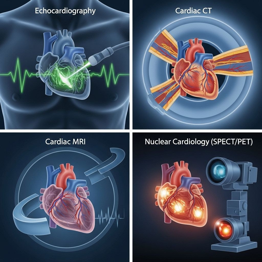
Non-Invasive Cardiac Imaging Comparison: Technologies, Diagnostic Value, and Clinical Impact
A clear view of each scan’s strengths and drawbacks to guide personalized heart care.

Non-Invasive Cardiac Imaging Comparison
Advancements in non-invasive cardiac imaging have revolutionized the diagnosis, risk stratification, and management of coronary artery disease (CAD). By providing reliable, patient-friendly alternatives to traditional invasive tests, modern imaging technologies facilitate timely and accurate clinical decision-making. This article comprehensively analyzes the evidence, strengths, limitations, and appropriate use scenarios for the main non-invasive cardiac imaging modalities, with special attention to their comparative diagnostic performance, utility, and clinical implications.
Table of Contents
- Introduction
- Key Non-Invasive Cardiac Imaging Technologies
- Diagnostic Comparisons: Sensitivity, Specificity, and Accuracy
- Clinical Applications and Patient Selection
- Advantages and Limitations of Each Modality
- Economic and Practical Considerations
- Future Directions and Innovations
- Frequently Asked Questions (FAQs)
Introduction
Cardiovascular disease represents the leading cause of mortality worldwide, with CAD being a primary driver of morbidity and mortality. The increasing burden of CAD necessitates accurate, efficient, and patient-centered diagnostic approaches. While invasive coronary angiography remains the reference standard for diagnosing significant coronary stenosis, non-invasive imaging modalities are indispensable for initial assessment, risk stratification, and therapeutic planning. Over the past two decades, technological developments have resulted in multiple sophisticated non-invasive options, including cardiac computed tomography angiography (CT Angio), magnetic resonance imaging (MRI), single-photon emission computed tomography (SPECT), and echocardiography, each offering unique diagnostic benefits.
Key Non-Invasive Cardiac Imaging Technologies
Major non-invasive cardiac imaging modalities in current clinical use include:
- Cardiac Computed Tomography Angiography (CCTA)
- Cardiac Magnetic Resonance Imaging (CMR)
- Single Photon Emission Computed Tomography (SPECT)
- Stress Echocardiography (ECHO)
Overview of Modalities
| Modality | Primary Application | Key Features |
|---|---|---|
| CCTA | Coronary anatomy, stenosis assessment, non-invasive angiography | High spatial resolution, rapid acquisition, visualizes coronary arteries |
| CMR | Perfusion imaging, myocardial viability, cardiac structure/function | Tissue characterization, functional analysis, no ionizing radiation |
| SPECT | Perfusion assessment | Widely available, functional assessment, uses radiotracers |
| Echocardiography | Wall motion, stress response, ejection fraction | Non-ionizing, portable, real-time imaging |
Brief Modality Descriptions
- Cardiac CT Angiography (CCTA): Utilizes multidetector CT scanners to visualize coronary anatomy, enabling assessment of plaque burden and luminal stenosis. Particularly useful for ruling out obstructive CAD in low-to-intermediate risk patients.
- Cardiac Magnetic Resonance Imaging (CMR): Employs powerful magnetic fields and radiofrequency pulses to generate highly detailed images, assessing myocardial perfusion, viability, edema, fibrosis, and cardiac morphology. CMR is pivotal for comprehensive tissue characterization.
- Single Photon Emission Computed Tomography (SPECT): A nuclear imaging modality that evaluates myocardial perfusion using radiotracers. SPECT provides functional information about ischemia but has lower spatial resolution than CCTA or CMR.
- Stress Echocardiography: Ultrasound-based assessment of wall motion abnormalities during exercise or pharmacologic stress. It evaluates myocardial function and can identify ischemia.
Diagnostic Comparisons: Sensitivity, Specificity, and Accuracy
When selecting an imaging test, clinicians consider diagnostic accuracy for detecting significant coronary artery disease. Comparative metrics include sensitivity, specificity, and area under the receiver operating characteristic curve (AUC).
Head-to-Head Performance Metrics
| Modality | Sensitivity (%) | Specificity (%) | Area Under Curve (AUC) |
|---|---|---|---|
| CCTA | 95 | 37 | 0.91 |
| CCTA + CT Perfusion (CTP) | 93 | 59 | 0.89 |
| CMR | 91 | 69 | 0.91 |
| SPECT | 63 | 66 | 0.70 |
- CCTA demonstrates the highest sensitivity but lower specificity, increasing the risk for false positives in low-prevalence populations.
- CCTA+CTP hybrid approach balances sensitivity with improved specificity versus CCTA alone.
- CMR provides both high sensitivity and moderate specificity, excelling in myocardial tissue assessment and viability.
- SPECT displays moderate specificity but lower sensitivity, resulting in potentially missed significant CAD cases, particularly in patients with multiple prior interventions or microvascular disease.
Summary Receiver Operator Characteristic (SROC) Curves
SROC curves visually summarize diagnostic test performance across studies. CCTA and CMR display the largest area under the curve (AUC ~0.91), indicating robust overall accuracy, while SPECT’s smaller AUC (0.70) reflects its limitations in sensitivity and diagnostic yield.
Clinical Applications and Patient Selection
The selection of an appropriate non-invasive imaging modality hinges on clinical indications, patient characteristics, and local expertise.
Common Clinical Scenarios
- Assessment of Suspected CAD: CCTA is often favored for rapid exclusion of obstructive disease in symptomatic, low-to-intermediate risk patients.
- Evaluation of Myocardial Viability: CMR is gold standard for viability and tissue characterization; PET is highly sensitive but resource intensive.
- Risk Stratification Before Invasive Procedures: SPECT or stress ECHO can be used for functional assessment, particularly in resource-limited settings.
- Complex Cases: Hybrid or multiparametric approaches (e.g., CCTA + CTP, CMR) may be necessary for patients with previous revascularization, prior myocardial infarction, or atypical presentations.
Patient-Centered Considerations
- Renal function (gadolinium or contrast contraindications)
- Radiation exposure (young patients or repeat imaging)
- Obesity or poor acoustic windows (limitations for ECHO)
- Ability to tolerate breath-holding or stress protocols
Advantages and Limitations of Each Modality
| Modality | Advantages | Limitations |
|---|---|---|
| CCTA |
|
|
| CMR |
|
|
| SPECT |
|
|
| Stress ECHO |
|
|
Economic and Practical Considerations
Cost-effectiveness and resource allocation shape imaging test selection. According to evidence-based analyses, PET is more sensitive and cost-effective compared to dobutamine echocardiography and SPECT, with less data available for direct comparison with CMR. Factors such as local availability, speed of test, and insurance coverage also influence routine practice. Portable modalities like ECHO and SPECT are common in community hospitals, while CMR and CCTA are typically housed in tertiary centers.
Future Directions and Innovations
- Hybrid Imaging: Integrates anatomical and functional data (e.g., CCTA + CTP, PET-MRI) for improved accuracy and risk assessment.
- Advanced Software & AI: Artificial intelligence is increasingly used for automated image interpretation, quantification, and risk modeling.
- Novel biomarkers & tracers: Development of radiotracers and techniques that better identify microvascular dysfunction, inflammation, or plaque instability.
- Radiation Dose Reduction: Lower-dose protocols and enhanced detector sensitivity diminish risk, especially important for repeated studies.
- Expanded Access: Efforts are underway to improve the availability and affordability of advanced modalities worldwide.
Frequently Asked Questions (FAQs)
Q: Which non-invasive modality is best for ruling out coronary artery disease?
A: CCTA is most effective for rapid exclusion due to its high sensitivity, especially in patients with low-to-intermediate risk.
Q: Which imaging test is preferred for assessing myocardial viability?
A: Cardiac MRI (CMR) and PET are leading choices, with CMR excelling in tissue characterization and PET demonstrating superior sensitivity.
Q: Is radiation a risk with all non-invasive cardiac imaging?
A: CCTA and SPECT both use ionizing radiation, while CMR and Echocardiography do not.
Q: How should imaging modality choice be tailored?
A: Test selection should consider patient risk factors, clinical presentation, contraindications (contrast media, renal failure, pacemakers), and resource availability.
Q: Are non-invasive imaging tests cost-effective?
A: Cost-effectiveness varies; PET has shown dominance over certain modalities but requires specialized resources. ECHO and SPECT remain more accessible and affordable for many institutions.
Conclusion
Non-invasive cardiac imaging continues to evolve, providing clinicians with an array of complementary techniques for diagnosis, risk stratification, and management of CAD. Selection of the appropriate modality depends on clinical indication, patient factors, and available infrastructure. When applied appropriately, these technologies promote optimal patient outcomes, minimize unnecessary invasive procedures, and facilitate precision cardiovascular care.
References
- https://www.ncbi.nlm.nih.gov/pmc/articles/PMC3377565/
- https://pmc.ncbi.nlm.nih.gov/articles/PMC3377536/
- https://pmc.ncbi.nlm.nih.gov/articles/PMC11671941/
- https://pmc.ncbi.nlm.nih.gov/articles/PMC5701553/
- https://healthmanagement.org/c/imaging/IssueArticle/non-invasive-cardiac-imaging-modalities
- https://www.aafp.org/pubs/afp/issues/2007/0415/p1219.pdf
- https://www.uscjournal.com/articles/advanced-non-invasive-imaging-techniques?language_content_entity=en
- https://www.radcliffecardiology.com/image-gallery/23447/14407/comparison-of-non-invasive-imaging-techniques
- https://pmc.ncbi.nlm.nih.gov/articles/PMC535457/
Read full bio of medha deb


