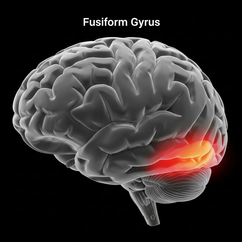
The Fusiform Gyrus: Neural Foundations of Vivid Visualization and Perception
Deep dive into how this brain region blurs the line between imagination and perception.

The human brain’s remarkable ability to conjure, differentiate, and recognize visual experiences—from faces and objects to vivid mental images—owes much to a specialized structure on its underside: the fusiform gyrus. Modern neuroscience has revealed this gyrus as a cornerstone of visual cognition, supporting both real and imagined visual experiences. This article examines the fusiform gyrus’s anatomy, its multifaceted role in visualization, object and word recognition, how it helps distinguish imagination from reality, and the clinical impacts of dysfunction in this region.
Table of Contents
- Introduction
- Anatomy and Structural Organization
- Core Functions of the Fusiform Gyrus
- Functional Subdivisions and Modular Organization
- Fusiform Gyrus and Vivid Visualization
- Distinguishing Reality from Imagination
- Clinical Relevance and Disorders
- Frequently Asked Questions (FAQs)
- Conclusion
Introduction
Visualization—the mental generation or re-experiencing of images beyond what is presently seen—enables us to imagine faces, recall scenic places, or mentally rehearse movements. The fusiform gyrus lies at the heart of this capability, connecting the flow of visual information from the eye to areas involved in recognition, memory, and even conscious discrimination between reality and imagination . This gyrus’s involvement ranges from perceiving faces and bodies to reading, with evidence suggesting that its activation intensity correlates with the vividness of mental visualization .
Anatomy and Structural Organization
The fusiform gyrus sits on the basal surface (underside) of the temporal and occipital lobes of the human brain. It is bordered medially by the parahippocampal gyrus and laterally by the inferior temporal gyrus, with the midfusiform sulcus dividing it into two main gyri:
- Medial occipitotemporal gyrus
- Lateral occipitotemporal gyrus
This location places it directly in the ventral visual processing stream (the “what” pathway) and grants it connectivity to primary and associative visual cortices as well as long-range connections to frontal and parietal lobes involved in higher cognition .
| Aspect | Description |
|---|---|
| Location | Basal surface of temporal and occipital lobes |
| Major Subdivisions | Medial and lateral occipitotemporal gyri (separated by midfusiform sulcus) |
| Key Connections | Primary/secondary visual cortex, angular gyrus, frontal and parietal lobes |
| Principal White Matter Tracts | Inferior longitudinal fasciculus, occipitotemporal association fibers |
Core Functions of the Fusiform Gyrus
The fusiform gyrus supports a variety of high-level visual and cognitive functions that underpin recognition and vivid mental imagery :
- Face recognition (including identifying individuals and decoding facial expressions)
- Visual word form processing (essential for reading)
- Body recognition and category-level object identification
- Color and shape integration
- Memory and multisensory integration
Its involvement in both sensory processing and higher-order tasks makes it a linchpin of multisensory perception and memory consolidation .
Key Specialized Regions within the Fusiform Gyrus
- Fusiform Face Area (FFA): Critical for recognizing individual faces and facial emotions. Damage in this area is closely associated with prosopagnosia (face blindness) .
- Visual Word Form Area (VWFA): Specialized for the recognition of written words, supporting fluent reading and distinguishing letters and letter strings .
- Body-selective Regions: Adjacent to the FFA, some regions are tuned for body form recognition, supporting the categorization of body shape and posture .
Functional Subdivisions and Modular Organization
Contemporary neuroimaging and tractography studies reveal that the fusiform gyrus is not a monolithic region but comprises distinct functional modules with specialized processing roles :
- Medial Fusiform Gyrus (FGm): Acts as a transition and integration zone, combining multiple stimuli types.
- Lateral Fusiform Gyrus (FGl): Encodes categorical recognition—particularly important for faces, objects, or bodies.
- Anterior Fusiform Gyrus (FGa): Enhances semantic processing and higher-order interpretation of visually derived information.
This modularity allows for both fine-grained visual discrimination—such as telling apart two nearly identical faces—and integration of complex sensory and semantic cues .
Fusiform Gyrus and Vivid Visualization
Visualization involves internally re-experiencing or synthesizing visual information in the absence of sensory input. Robust evidence demonstrates that the fusiform gyrus is intensely active during tasks that require participants to imagine or recall detailed images, scenes, or objects.
- Functional MRI studies show that greater activation of the fusiform gyrus corresponds to the vividness and clarity of mental imagery .
- Those with a highly responsive fusiform gyrus are capable of forming more detailed and lifelike visualizations.
- Imagining faces, reading words in one’s mind, or recalling visual scenes all recruit this region, often mirroring neural activity patterns seen during actual perception .
Mental imagery therefore draws on the same neural machinery that supports direct perception, explaining why some imagined experiences can feel intensely real and detailed .
Mechanisms Underlying Vivid Visualization
- Top-Down Modulation: Higher-order regions (e.g., prefrontal cortex) can stimulate the fusiform gyrus, triggering imagery even in the absence of external visual input.
- Overlap with Reality: The similarity of activation patterns in perception and visualization blurs the boundary between image and reality in the mind’s eye.
- Variation Across Individuals: People differ in their baseline fusiform responsiveness, accounting for differences in the vividness of inner imagery (some can form photorealistic visuals, others only vague shapes or outlines).
Distinguishing Reality from Imagination
One of the brain’s subtler functions is its ability to distinguish real sensory experiences from imaginary ones. Disruptions to this faculty are linked to hallucinations or mistaken beliefs about reality (as seen in some psychiatric disorders). Recent studies highlight:
- Strong fusiform gyrus activation during vivid mental imagery increases the chance a person will mistake an internal image for something actually seen .
- A network including the fusiform gyrus and anterior insula helps the brain “tag” experiences as real or imagined. When these systems falter, boundaries blur between imagination and perception.
- This distinction is crucial not only for healthy cognition but also has direct implications for conditions such as schizophrenia or visual hallucinations.
Key Research Insight: When individuals are asked to vividly imagine a patterned visual scene, brain scans reveal that the fusiform gyrus lights up almost as strongly as if the scene had been presented on a screen. Those with particularly vivid imagery—and especially those whose anterior insula activity is low—are most likely to confuse their own mental images for reality .
Role in Imagination vs. Reality Table
| Feature | Imagination | Reality |
|---|---|---|
| Fusiform Gyrus Activation | High (vivid imagery) | High (vivid perception) |
| Anterior Insula Involvement | Critical for reality discrimination | Acts as comparator with incoming signals |
| Risk of Confusion | Increases with intense imagery, weak insula tagging | Usually low, unless pathological |
Clinical Relevance and Disorders
Damage, dysfunction, or developmental anomalies in the fusiform gyrus have wide-ranging cognitive and perceptual consequences:
- Prosopagnosia (Face Blindness): Injury or underdevelopment of the FFA leads to impaired recognition of faces despite normal vision .
- Dyslexia: Disruption of the VWFA can result in reading and letter recognition difficulties.
- Synesthesia: Aberrant fusiform connectivity may contribute to blending of sensory experiences, such as seeing colors when reading specific words or numbers .
- Visual Hallucinations and Schizophrenia: Excessive fusiform gyrus activation and breakdown of reality tagging mechanisms can cause the brain to misinterpret imagined images as real .
- Alzheimer’s Disease and Dementia: Early dysfunction in this zone may underlie visual misperceptions or memory-related recognition deficits.
Because the fusiform gyrus underpins both the richness of mental imagery and the ability to correctly identify and interpret visual stimuli, its healthy function is essential for normal cognition and perception.
Frequently Asked Questions (FAQs)
Q: What is the fusiform gyrus?
A: The fusiform gyrus is a spindle-shaped brain region on the underside of the temporal and occipital lobes, involved in high-level visual processing, including face, word, and object recognition as well as memory and vivid visualization.
Q: How does the fusiform gyrus contribute to mental imagery?
A: It helps recreate detailed visual scenes or objects in the absence of actual sensory input, with the degree of activation influencing how vivid and realistic the formed image feels.
Q: Why do some people have more vivid visualizations than others?
A: Variability in the responsiveness and connectivity of an individual’s fusiform gyrus, along with interactions with prefrontal and insular regions, affects the vividness of internal imagery.
Q: What happens if the fusiform gyrus is damaged?
A: Damage can cause difficulties with facial recognition (prosopagnosia), reading (if the visual word form area is affected), object recognition, or increased risk of visual hallucinations and confusion between imagination and reality.
Conclusion
The fusiform gyrus is a neural powerhouse enabling humans to navigate the visual world, imagine richly detailed scenarios, and seamlessly identify objects, words, and faces. Its unique role in bridging perception and visualization makes it central to imaginative thought, creative visualizations, and essential everyday recognitions. Understanding how this gyrus operates—both when it functions seamlessly and when its processes go awry—provides critical insight into human cognition, the nature of reality and imagination, and the future of neurocognitive research.
References
- https://www.kenhub.com/en/library/anatomy/fusiform-gyrus
- https://pmc.ncbi.nlm.nih.gov/articles/PMC6867330/
- https://neurosciencenews.com/reality-imagination-brain-29221/
- https://radiopaedia.org/articles/fusiform-gyrus?lang=us
- https://www.nature.com/articles/s41598-020-70410-6
- https://www.droracle.ai/articles/60018/fusiform-gyrus-brain
- https://en.wikipedia.org/wiki/Visual_word_form_area
Read full bio of Sneha Tete


