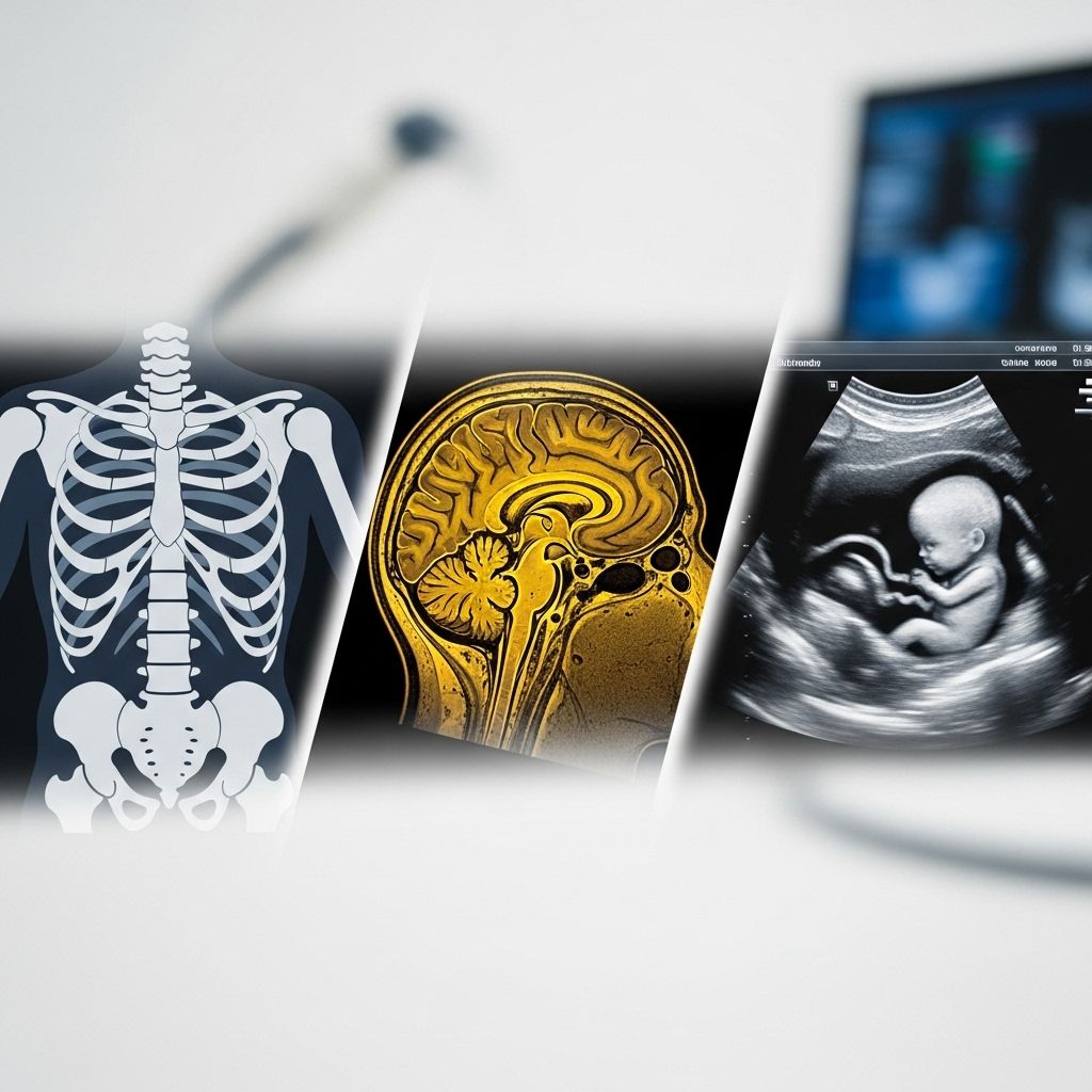
Diagnostic Imaging Fundamentals: Understanding X-Ray, MRI, and CT Scan Technologies
Understand how each imaging technique works and its optimal use in patient care.

Diagnostic Imaging Basics: X-Ray, MRI, CT Scan
Modern medicine relies heavily on diagnostic imaging to investigate and diagnose a wide range of conditions. Among the most widely used imaging modalities are X-rays, Magnetic Resonance Imaging (MRI), and Computed Tomography (CT) scans. Each technology has unique principles, uses, strengths, and safety considerations. This comprehensive guide introduces and compares these core diagnostic tools, demystifying their differences and practical applications for patients and healthcare professionals alike.
Table of Contents
- Introduction to Diagnostic Imaging
- What Is an X-Ray?
- What Is a CT Scan?
- What Is an MRI?
- Comparison Chart: X-Ray vs. CT vs. MRI
- Choosing the Right Imaging Method
- Frequently Asked Questions (FAQs)
Introduction to Diagnostic Imaging
Diagnostic imaging encompasses a suite of technologies that generate visual representations of the interior of the body for clinical analysis. These images are crucial in identifying abnormalities, confirming diagnoses, guiding treatments, and monitoring disease progression.
Among the most prevalent modalities are:
- X-rays — Simple, quick 2D images developed using ionizing radiation.
- CT Scans — Advanced, computer-processed X-rays that construct detailed 3D images.
- MRIs — Radiation-free technique utilizing strong magnets and radio waves for highly detailed images, especially of soft tissues.
Understanding how each technology works, their best applications, and unique cautions helps both providers and patients make informed decisions.
What Is an X-Ray?
X-rays, discovered in the late 19th century, were the first and remain the simplest form of diagnostic imaging. X-ray machines emit a controlled beam of ionizing radiation that passes through the body, with varying absorption by different tissues producing a contrast image on detector film or digital sensors.
How X-Rays Work
- The machine directs a stream of ionizing radiation through the body.
- Dense structures (like bones and teeth) absorb more radiation, appearing white on the image.
- Softer tissues (like flesh and organs) absorb less, appearing darker.
- The result is a two-dimensional projection highlighting key anatomical differences.
Common Uses of X-Rays
- Bone fractures and dislocations
- Chest conditions (e.g., detecting pneumonia, fluid in lungs, or tumors)
- Dental assessments (teeth, jawbone health, cavities)
- Detecting certain tumors and foreign objects
X-rays are usually the first imaging method chosen in emergency rooms and clinics due to their speed and accessibility.
Advantages and Limitations of X-Rays
- Rapid and widely available
- Low cost compared to other modalities
- Excellent for imaging bones and dense tissues
- Limited in visualizing soft tissue, ligaments, tendons
- Cannot produce 3D images; only 2D projections
Safety and Risks of X-Rays
- X-rays use ionizing radiation, which can damage cells and DNA with excessive or repeated exposure
- Single diagnostic exposures are generally considered very low risk
- Special caution required for pregnant women and children
What Is a CT Scan?
Computed Tomography (CT) scans are an advanced form of X-ray imaging that enables highly detailed, three-dimensional visualization of internal anatomy. Sometimes called a “CAT scan,” a CT utilizes computer processing to combine multiple X-ray measurements taken from different angles.
How CT Scans Work
- A rotating X-ray tube circles the patient, capturing multiple 2D images (“slices”) at successive angles
- A computer reconstructs these slices into a composite 3D cross-sectional image
- Contrast agents may be introduced to better visualize specific tissues or blood vessels
Common Uses of CT Scans
- Emergency trauma (detecting internal bleeding, organ injury, bone fractures)
- Identifying tumors and cancers
- Assessing blood vessels (e.g., pulmonary embolism, aneurysms)
- Guidance for biopsies or surgical planning
CT scans are more comprehensive than basic X-rays and are often ordered when more anatomical detail is needed.
Advantages and Limitations of CT Scans
- Rapid scan times (entire areas can be imaged in minutes)
- Cross-sectional (3D) imaging for precise diagnosis
- Better visualization of organs, blood vessels, soft tissue than standard X-rays
- Not as detailed as MRI for certain soft tissue and neural structures
- More radiation exposure than a basic X-ray
Safety and Risks of CT Scans
- Significant exposure to ionizing radiation in comparison with X-rays
- Potential allergic reaction to contrast agents
- Generally not advised for pregnant women unless absolutely necessary
- Potential, though small, increased risk of cancer with heavy or repeated scans
What Is an MRI?
Magnetic Resonance Imaging (MRI) is a breakthrough technology for detailed visualization of both hard and soft tissues—without the use of ionizing radiation. Instead, MRI relies on strong magnetic fields and radio frequency pulses to generate images, making it uniquely valuable for evaluating subtle differences between types of tissues.
How MRIs Work
- The patient enters a large, cylindrical magnet
- Radio waves are pulsed through the body, causing hydrogen atoms within the tissue to align and resonate
- Signals emitted by the atoms are captured and processed into high-resolution images
- The computer arranges the data into detailed cross-sectional slices or 3D reconstructions
Common Uses of MRIs
- Soft tissue evaluation (brain, spinal cord, joints, muscles, ligaments, tendons)
- Certain cancers (particularly those affecting the brain or soft tissue)
- Neurological disorders (multiple sclerosis, stroke, tumors)
- Cardiac and blood vessel imaging (magnetic resonance angiography/MRA)
MRIs are the gold standard for non-invasive imaging of the central nervous system and most musculoskeletal disorders.
Advantages and Limitations of MRIs
- No radiation exposure
- Superior visualization for soft tissues, neural structures, joint components
- Useful for detecting subtle or early-stage abnormalities
- Longer scan times (often 30 minutes or more)
- Cannot be used in patients with metal implants, pacemakers, or certain foreign objects
- More expensive and less widely available than X-rays or CT scans
Safety and Risks of MRIs
- Considered very safe for most patients
- No exposure to radiation
- Risks for patients with metal implants (can heat, move, or malfunction in strong magnetic fields)
- Patients may experience claustrophobia or anxiety within the MRI tunnel
- Sensitive to patient movement, which can blur images
Comparison Chart: X-Ray vs. CT vs. MRI
| Feature | X-Ray | CT Scan | MRI |
|---|---|---|---|
| Basic Principle | Ionizing radiation (2D image) | Multiple X-rays + computer processing (3D image) | Strong magnets & radio waves (3D image) |
| Radiation Exposure | Low | Higher | None |
| Image Detail | Bone, some soft tissues | Bone, organs, soft tissue | Excellent for soft tissues, nerves, brain |
| Scan Time | Minutes | Minutes | 30-60 minutes |
| Common Uses | Bone fractures, chest, dental | Trauma, cancers, organs | Brain, nerves, joints, soft tissue |
| Risks | Radiation/ Pregnancy caution | Higher radiation/Contrast allergies | Claustrophobia/Metal implant risk |
| Cost | Least expensive | Moderate | Most expensive |
Choosing the Right Imaging Method
The selection of diagnostic imaging technique depends on a combination of factors:
- Type of tissue or injury (bone fractures: X-ray or CT; soft tissue injury: MRI)
- Urgency (CT for rapid trauma assessment, X-ray for immediate bone injury)
- Patient safety (avoid MRI with unsafe metal implants, minimize radiation in pregnancy)
- Need for anatomical detail (MRI for nerve or ligament analysis, CT for full organ cross-sections)
- Clinical guidelines and cost
Healthcare providers balance these aspects to optimize diagnostic yield while limiting risk.
Frequently Asked Questions (FAQs)
Q: Which scan is safest: X-ray, CT, or MRI?
A: MRI is generally the safest as it uses no ionizing radiation. X-rays and CT scans are also safe for most when used judiciously, but both use radiation, with CT exposure much higher than X-ray.
Q: Why do some people need a CT after an X-ray?
A: CT scans provide a more detailed, three-dimensional view of complex or ambiguous findings seen on an X-ray, especially for internal organs or subtle bone injuries.
Q: Is MRI always better than CT or X-ray?
A: Not always. MRI excels at soft tissue and neural imaging, but CT is preferred for acute trauma, complex fractures, or patients for whom MRI is contraindicated (e.g., due to metal implants).
Q: Can I have these scans while pregnant?
A: MRI (without contrast) is considered safe in pregnancy when truly necessary. X-rays and CT scans are generally avoided unless emergency evaluation is required due to potential fetal radiation exposure. Always inform your provider if you are or may be pregnant.
Q: Are contrast agents used in all scans?
A: No. Contrast agents are only used when greater differentiation between tissues is needed (e.g., highlighting blood vessels, tumors), and are more common in CT and MRI than in X-rays. Inform your doctor of any allergies or kidney problems before receiving contrast.
Key Takeaways
- X-rays are fast, low-cost, 2D, and best for bones.
- CT scans offer rapid 3D visualization ideal for trauma and internal organs, but at the cost of higher radiation.
- MRIs provide superior soft-tissue detail using magnets, with no radiation, but are more expensive, take longer, and are sensitive to metal.
Selecting the proper imaging technique is individualized. Work with your healthcare provider to determine the safest and most informative approach for your situation.
References
- http://www.wth.org/blog/ct-scans-mris-and-x-rays-oh-my-making-sense-of-imaging/
- https://www.envrad.com/difference-between-x-ray-ct-scan-and-mri/
- https://www.premierortho.org/the-difference-between-x-ray-ct-and-mri/
- https://www.healthline.com/health/ct-scan-vs-mri
- https://orthoinfo.aaos.org/en/treatment/x-rays-ct-scans-and-mris/
- https://pmc.ncbi.nlm.nih.gov/articles/PMC9192206/
- https://singingriverhealthsystem.com/2022/12/radiology-for-dummies/
- https://www.kenhub.com/en/library/anatomy/medical-imaging-and-radiological-anatomy
Read full bio of medha deb


