Upper GI Endoscopy: Procedure, Preparation, and Recovery
Understand the complete process, indications, preparation, and recovery involved in upper GI endoscopy.

Upper gastrointestinal (GI) endoscopy, also known as esophagogastroduodenoscopy (EGD), is a vital diagnostic and therapeutic tool used to examine the inside of the upper digestive tract. This minimally invasive procedure gives physicians a direct view of the esophagus, stomach, and duodenum, assisting in identifying and sometimes treating underlying digestive tract disorders.
What Is an Upper GI Endoscopy?
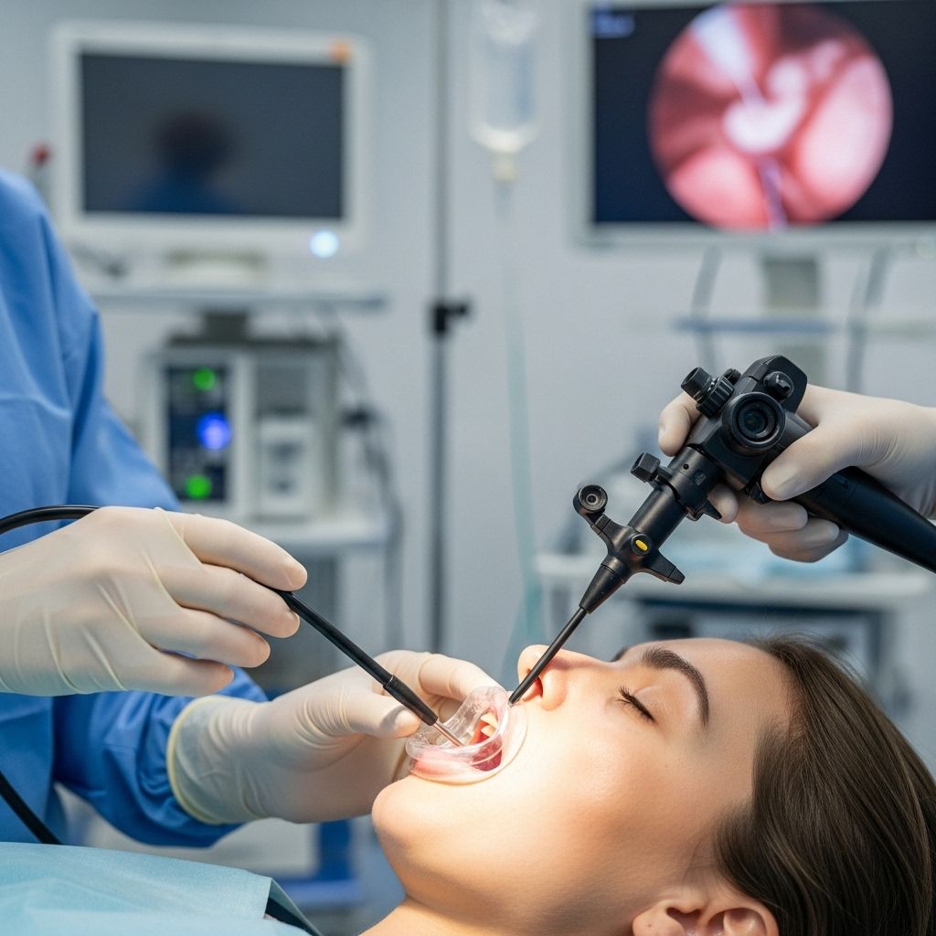
Upper GI endoscopy is a procedure that allows doctors to visualize the interior of the upper digestive system—namely the esophagus, stomach, and the first part of the small intestine (duodenum). It utilizes an endoscope, which is a long, thin, flexible tube equipped with a tiny camera and light source at its tip. Sometimes, this procedure is also referred to as an esophagogastroduodenoscopy (EGD).
- Purpose: Diagnosing and sometimes treating problems of the upper digestive tract.
- Device: Flexible endoscope inserted through the mouth.
- Usual Setting: Hospital or outpatient center.
Why Is an Upper GI Endoscopy Performed?
There are several reasons your healthcare provider may recommend an upper GI endoscopy:
- Evaluation of Symptoms: Investigating causes of symptoms such as persistent upper abdominal pain, nausea, vomiting, heartburn, trouble swallowing, or unexplained weight loss.
- Diagnosis: Detection of ulcers, tumors, inflammation, infection, or bleeding within the digestive tract.
- Biopsies: Taking tissue samples to test for infections (like Helicobacter pylori), celiac disease, cancer, or other abnormalities.
- Treatment: Directly addressing issues such as stopping active bleeding, removing polyps or foreign objects, dilating narrowed sections (strictures), or treating abnormal tissue such as a tumor using specialized endoscopic tools.
- Surveillance: Monitoring the progression of chronic conditions, such as Barrett’s esophagus or gastric ulcers.
Conditions Diagnosed or Treated by Upper GI Endoscopy
The procedure can help detect or manage:
- Esophagitis and gastroesophageal reflux disease (GERD)
- Stomach (gastric) or duodenal ulcers
- Benign or malignant (cancerous) tumors of the esophagus, stomach, or duodenum
- Barrett’s esophagus
- Hiatal hernia
- Strictures (narrowing of the esophagus or pylorus)
- Varices (enlarged veins, especially in liver disease)
- Celiac disease
- Unexplained upper GI bleeding
How to Prepare for an Upper GI Endoscopy
Proper preparation increases the accuracy and safety of the procedure.
- Fasting: You will need to stop eating and drinking for at least 6 to 8 hours before the test. This ensures the stomach and duodenum are empty and can be seen clearly.
- Medications: Inform your doctor about all medicines and supplements. You may need to adjust or stop certain medications such as blood thinners, antiplatelets, or diabetes medications.
- Allergies and Medical Conditions: Tell your healthcare team about allergies, bleeding disorders, or heart and lung conditions.
- Consent: Before the procedure, you will review and sign a consent form after discussing risks and benefits with your provider.
- Transportation: Because sedation is commonly used, arrange for someone to take you home afterward.
Preparation Checklist
- Follow specific fasting instructions given by your doctor.
- Discuss which medications you should withhold or adjust.
- Prepare a list of current medications, allergies, and medical history for your care team.
- Arrange post-procedure transportation and, if needed, time off from work.
What Happens During the Procedure?
The steps and experience during an upper GI endoscopy typically include:
- Arrival and Preparation
- Review of your health history and vital sign check.
- Change into a hospital gown.
- Insertion of an intravenous (IV) line for sedation.
- Sedation and Anesthesia
- Medication to relax and make you drowsy (conscious sedation) or occasionally general anesthesia.
- Local anesthetic sprayed on your throat to numb the area and minimize gag reflex.
- Insertion of the Endoscope
- You will lie on your left side on the exam table.
- Placement of a mouthguard or bite block to protect teeth and the endoscope.
- Doctor gently guides the lubricated endoscope through the mouth, down the throat, and into the esophagus, stomach, and duodenum.
- The device does not interfere with breathing.
- Visualization and Procedures
- The camera sends live images to a video monitor as the physician examines the tissue lining.
- If necessary, the doctor can perform biopsies, remove polyps, cauterize bleeding vessels, dilate narrowed areas, or remove objects using specialized tools passed through the endoscope.
- Completion and Recovery
- The endoscope is slowly withdrawn.
- You are moved to a recovery area as the sedation wears off. Most people rest and wake comfortably within an hour.
- Bloating, mild sore throat, or gas may occur temporarily after the test.
Key Features of the Endoscope
- Flexible and thin (about the size of a little finger)
- Embedded with a light and high-definition video camera
- Channels that allow instruments to be inserted for biopsy or treatment
Risks and Possible Complications
Upper GI endoscopy is a generally safe procedure, but all medical tests come with some risks. These are uncommon but should be considered:
- Bleeding, especially with biopsies or removal of polyps
- Perforation (a small tear in the esophagus, stomach, or duodenum lining)
- Infection (rare, especially if no tissue is removed)
- Reaction to sedation or anesthesia
- Temporary sore throat, hoarseness, or dental injury from insertion
- Heart or lung complications (rare, linked to sedation in those with pre-existing problems)
Your medical team will discuss specific risks based on your health status before the procedure.
What to Expect After Upper GI Endoscopy
- You will be observed until effects of sedation wear off—generally 30 minutes to 2 hours.
- Temporary symptoms may include sore throat, grogginess, mild bloating, or hunger.
- Do not eat or drink until swallowing improves and numbness from anesthetic spray passes.
- Avoid driving, operating machinery, or making important decisions for the remainder of the day due to lingering sedation.
- Your doctor will explain initial findings; biopsy and sample results may take several days.
- Contact your doctor if you experience severe pain, persistent vomiting, difficulty breathing, high fever, or black/tarry stools after the procedure.
Benefits of Upper GI Endoscopy
- Direct visualization of the upper digestive tract.
- Ability to take targeted biopsies under direct sight.
- Simultaneous diagnostic and therapeutic interventions.
- Generally safe, outpatient, and well-tolerated.
- Quicker recovery and lower complication rates compared to surgical approaches for certain issues.
Alternatives to Upper GI Endoscopy
Depending on the situation, your physician may discuss alternative options, such as:
- Barium swallow X-rays: Evaluates swallowing and the structure of the esophagus and upper stomach.
- Capsule endoscopy: Patient swallows a tiny camera in a pill to take pictures as it travels through the digestive tract (less targeted and no intervention possible during exam).
- CT or MRI scans: Advanced imaging, but without direct biopsy or treatment capability.
However, upper GI endoscopy offers both direct visualization and the opportunity to obtain tissue or provide therapy—making it the preferred first-line tool in many cases.
Frequently Asked Questions (FAQs) About Upper GI Endoscopy
Q: Will I be asleep during the procedure?
A: Most patients receive sedative medication and are relaxed or drowsy but can usually respond to instructions. Some may have deeper anesthesia if required, especially children or those with complex medical needs.
Q: Does the procedure hurt?
A: The procedure is generally not painful. Sedation and throat numbing minimize discomfort. Some pressure or bloating may be felt as air is introduced for better visualization.
Q: How long does an upper GI endoscopy take?
A: The examination itself usually takes 10–30 minutes, but expect to spend a few hours at the facility, including preparation and recovery time.
Q: Can I drive home after my endoscopy?
A: No. Sedative effects can impair reaction time and judgment for several hours. Someone else should drive you home and be with you for the remainder of the day.
Q: Are there any special risks if I have heart disease or another chronic condition?
A: Your doctor will assess your risks based on your medical background and may coordinate with your other healthcare providers to optimize safety. Always provide a full, up-to-date medication and allergy list before the procedure.
Q: How soon will I get results?
A: Preliminary findings are often discussed after recovery. Biopsy results or special studies may take several days to return, at which point your doctor will explain the findings and outline next steps if needed.
Q: What symptoms should prompt an urgent call to my doctor after the test?
A: Call your healthcare provider immediately for:
- Severe or persistent abdominal pain
- Black, tarry, or bloody stools
- Fever over 101°F (38.3°C)
- Shortness of breath, chest pain, or trouble swallowing
- Uncontrolled vomiting, especially if it is bloody or looks like coffee grounds
Summary Table: Key Points About Upper GI Endoscopy
| Aspect | Key Details |
|---|---|
| Definition | Minimally invasive exam of esophagus, stomach, duodenum using a flexible tube with a camera |
| Other Names | Esophagogastroduodenoscopy (EGD) |
| Purpose | Diagnose, biopsy, or treat upper GI symptoms/problems |
| Preparation | Fasting, medication adjustment, consent review |
| Sedation | Typically conscious sedation; anesthesia as needed |
| Procedure Duration | 10–30 minutes (plus pre- and post-procedure time) |
| Common Risks | Mild sore throat, bleeding, perforation, infection, sedation reactions |
| Alternatives | Barium swallow, capsule endoscopy, imaging (CT/MRI) |
Conclusion
Upper GI endoscopy is a safe, effective procedure for diagnosing and managing a range of upper digestive system issues. With clear preparation steps, skilled medical teams, and minimal downtime, this test remains a cornerstone of modern gastrointestinal medicine.
References
- https://www.niddk.nih.gov/health-information/diagnostic-tests/upper-gi-endoscopy
- https://www.cancer.org/cancer/diagnosis-staging/tests/endoscopy/upper-endoscopy.html
- https://patient.gastro.org/upper-gi-endoscopy/
- https://my.clevelandclinic.org/health/procedures/22549-egd-procedure-upper-endoscopy
- https://www.healthdirect.gov.au/surgery/upper-gi-endoscopy-and-colonoscopy
- https://www.youtube.com/watch?v=OmsNjUOvbSs
- https://healthy.kaiserpermanente.org/health-wellness/health-encyclopedia/he.upper-gastrointestinal-gi-endoscopy.hw267678
Read full bio of Sneha Tete


