Transesophageal Echocardiogram: Procedure, Preparation, and Insights
Learn the procedure, preparation steps, uses, risks, and frequently asked questions about transesophageal echocardiogram (TEE) imaging.

Transesophageal Echocardiogram (TEE): Comprehensive Guide
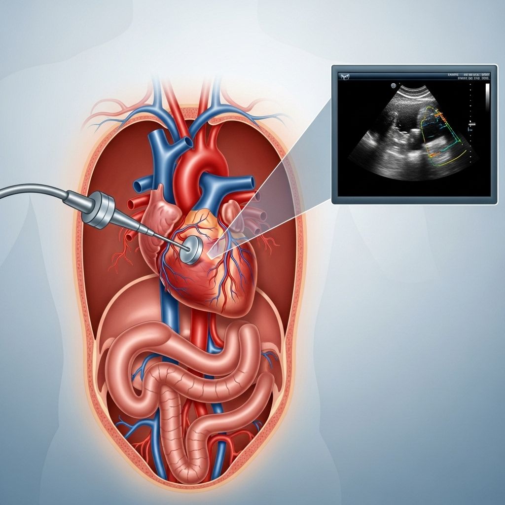
A transesophageal echocardiogram (TEE) is an advanced diagnostic imaging technique used to obtain exceptionally clear and detailed images of the heart and its structures. Unlike a traditional transthoracic echocardiogram, which is performed externally, a TEE involves guiding an ultrasound probe down the esophagus to get closer to the heart. This unique approach allows healthcare professionals to assess various heart conditions with greater precision.
What is a Transesophageal Echocardiogram?
A TEE is a special form of echocardiogram that uses high-frequency sound waves (ultrasound) to generate real-time images of the heart and adjacent blood vessels. By placing a transducer in the esophagus rather than on the chest, it helps obtain images with less interference from bone and lung tissue, leading to higher image quality and more accurate diagnoses.
- Transducer placement: The probe is guided through the mouth and positioned in the esophagus, which sits behind the heart.
- Clarity: Because of its proximity, the TEE provides vivid, comprehensive images of cardiac structures, especially those at the back of the heart.
- Purpose: Used to detect blood clots, evaluate heart valves, assess for infections, and guide heart procedures.
Why is a Transesophageal Echocardiogram Done?
The TEE is performed to provide a closer and more detailed look at the heart. This is particularly valuable when:
- Standard transthoracic echocardiogram (TTE) images are inconclusive or insufficiently detailed.
- Evaluating heart valve disorders, such as stenosis or regurgitation.
- Identifying blood clots in the heart chambers or attached to artificial valves.
- Assessing the presence of infective endocarditis (infection of heart tissues or valves).
- Investigating congenital heart defects, tumors, or masses within the heart.
- Guiding treatments for arrhythmias or cardiac surgeries.
- Examining the thoracic aorta for aneurysms, dissections, and other pathologies.
Common Indications for TEE
| Condition | Reason for TEE |
|---|---|
| Stroke or TIA | Look for clots or sources of emboli in the heart |
| Heart Valve Disease | Detailed evaluation of valve structure and function |
| Suspected Endocarditis | Detect vegetations (infective growths) on valves |
| Congenital Heart Defect | Visualize shunts, septal defects, or abnormal connections |
| Cardiac Tumor or Mass | Identify and assess size, location, and impact |
| Aortic Disease | Evaluate aortic dissection or aneurysm |
Preparing for a Transesophageal Echocardiogram
Proper preparation is vital to ensure the safety and effectiveness of a TEE procedure. Below are preparation steps typically advised by healthcare providers:
- Fasting: Do not eat or drink for at least 4–6 hours prior to the test. This precaution reduces the risk of aspiration when the probe is inserted.
- Medication: Take your usual prescribed medications with a minimal sip of water, unless directed otherwise by your provider.
- Transportation: Arrange for someone to drive you home as sedatives used during the test may cause lingering drowsiness.
- Allergy and Health History: Inform your healthcare provider about any allergies, medical conditions, problems swallowing, esophageal disease, or if you are on blood thinners.
- Anticoagulation: Notify staff if you are using anticoagulant (blood thinning) medications.
What to Expect During the TEE Procedure
The TEE procedure typically follows a well-defined process to ensure patient safety and comfort:
- Pre-Procedure: You will be asked to remove dentures, glasses, and jewelry. An intravenous (IV) line will be placed for sedation. Oxygen may be administered through a nasal cannula if needed.
- Anesthesia: The back of your throat will be numbed using a local anesthetic spray or gel to minimize discomfort during probe insertion.
- Sedation: A mild sedative is administered intravenously to help you relax and minimize gagging or anxiety. Typically, you remain awake but drowsy and able to follow instructions.
- Probe Insertion: A thin, flexible tube (roughly the size of an index finger) with an ultrasound transducer at its tip is gently inserted through your mouth and advanced into the esophagus.
- Imaging: The transducer emits ultrasound waves; echoes reflect off heart structures and are processed by a computer into moving images displayed on a monitor.
- Adjustments: The physician manipulates the probe to obtain images from various angles, thoroughly examining heart valves, chambers, vessels, and nearby tissues.
- Duration: The image acquisition phase usually takes 15–30 minutes, though the entire appointment may last up to 90 minutes including sedation and recovery time.
- Post-Procedure: Once complete, the probe is withdrawn and you are monitored until the effects of sedation wear off.
What You Might Feel
- Mild discomfort or gagging when the probe is inserted (reduced by numbing the throat and sedation).
- Pressure or slight movement in the throat as the probe is manipulated.
- You should not experience pain or difficulty breathing. Breathing is not blocked at any time during the procedure.
After the Procedure
Recovery from a TEE is generally quick and straightforward. Here’s what to expect:
- Observation for a short period until sedation wears off and you can swallow normally.
- Temporary sore throat or mild hoarseness may occur but typically resolves within a day.
- No eating or drinking (including water) for about 30–60 minutes post-procedure or until your gag reflex returns, to prevent choking.
- Avoid driving, operating machinery, or making important decisions for the next 24 hours due to sedation effects.
- Arrange for a friend or family member to accompany you home.
Risks and Possible Complications
While TEE is considered a safe test, it is more invasive than standard ultrasound and carries certain risks. Fortunately, serious complications are rare. Possible risks include:
- Sore throat: Most common, usually mild and short-lived.
- Minor bleeding: From irritation or minor trauma to the esophagus or throat lining.
- Injury: Rare risk of esophageal tear or perforation, especially in patients with esophageal disease or varices.
- Reactions to sedation: Including breathing difficulties or allergic reactions (uncommon with careful monitoring).
- Arrhythmias: Rare but possible, especially in people with pre-existing heart conditions.
- Aspiration: Low risk of inhaling food or fluids into the lungs if proper fasting is not followed.
Inform your provider immediately if you experience difficulty swallowing, chest pain, persistent sore throat, fever, or bleeding following the test.
TEE vs. Transthoracic Echocardiogram (TTE)
| Feature | TEE | TTE |
|---|---|---|
| Imaging Method | Probe in esophagus (internal) | Probe on chest wall (external) |
| Clarity of Images | Very high (especially for posterior structures) | Moderate, may be limited by bone/lung tissue |
| Invasiveness | Invasive | Noninvasive |
| Sedation Required | Yes | No (usually) |
| Used When | Deeper evaluation needed, TTE inconclusive | First-line screening |
| Patient Comfort | May cause throat discomfort | Generally comfortable |
Understanding Your TEE Results
The images and findings from your TEE are interpreted by a cardiologist or a cardiac imaging specialist. They provide a detailed report that covers:
- Heart anatomy and structural details
- Valve structure and function (e.g., leakage or stenosis)
- Presence of blood clots, tumors, or infections
- Abnormalities of the aorta
- Congenital defects or shunts
- General heart function and pumping ability
Your provider will discuss the findings and next steps. Results are usually available within 1–2 business days, but urgent discoveries may prompt immediate action or follow-up care.
Frequently Asked Questions (FAQs)
What makes TEE images more detailed than standard echocardiograms?
Because the probe is closer to the heart and not blocked by ribs or lungs, a TEE provides much clearer and more detailed images compared to a standard (transthoracic) echocardiogram.
Is a TEE painful?
Most patients experience little to no pain. The throat is numbed and sedation is provided for comfort. Some mild pressure or gagging might be felt when the probe is inserted, but the test does not interfere with breathing.
How long does the TEE test take?
The test itself typically takes 20 to 40 minutes, though preparing and recovering can mean you spend 1-2 hours in the clinic.
Can you drive after having a TEE?
No. The sedatives used can make you drowsy and affect coordination. You’ll need someone to drive you home, and you should not drive for at least 24 hours after the procedure.
Does a TEE test require general anesthesia?
It rarely requires general anesthesia. Most people are comfortably relaxed with mild to moderate sedation.
Who should not get a TEE?
Patients with severe esophageal disorders, such as strictures, tears, or active bleeding, may not be suitable for TEE. Your provider will evaluate your individual risks prior to recommending the test.
How soon will I get my results?
Most centers provide results to the patient’s referring doctor within a few days. Urgent findings will be shared sooner when clinically necessary.
Summary: Key Takeaways about Transesophageal Echocardiogram
- TEE is a specialized ultrasound of the heart performed via the esophagus for high-resolution images.
- It is useful for assessing valves, clots, infections, tumors, and structural abnormalities.
- Requires pre-test fasting, sedation, and post-test observation.
- Risks are low and complications rare, but the procedure is more invasive than standard echo.
- Results help inform diagnosis and management of a wide range of heart conditions.
If you have concerns or need to undergo a transesophageal echocardiogram, discuss your questions and medical history openly with your provider for the safest, most effective care experience.
References
- https://www.heartandstroke.ca/heart-disease/tests/transesophageal-echocardiogram-tee
- https://my.clevelandclinic.org/health/diagnostics/4992-echocardiogram-transesophageal-tee
- https://cardiology.weillcornell.org/sites/default/files/downloads/Guide%20to%20Transesophageal%20Echocardiography.pdf
- https://stanfordhealthcare.org/medical-tests/t/transesophageal-echocardiogram.html
- https://www.asecho.org/wp-content/uploads/2014/05/2013_Performing-Comprehensive-TEE.pdf
- https://www.ncbi.nlm.nih.gov/books/NBK442026/
- https://www.youtube.com/watch?v=iko2FZ1R9Ow
- https://www.massgeneral.org/heart-center/treatments-and-services/transesophageal-echocardiogram
- https://drsono.com/blogs/news/transesophageal-echocardiogram-tee-explained-see-details/
Read full bio of medha deb


