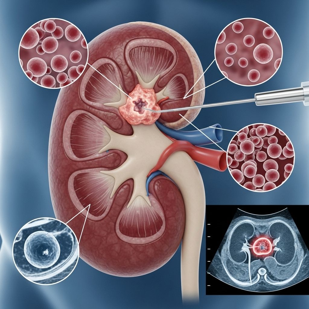
Testing for Renal Cell Carcinoma: Diagnosis, Approaches, and What to Expect
A comprehensive guide to diagnostic methods, imaging tests, and what to expect during renal cell carcinoma testing.

Understanding how renal cell carcinoma (RCC), the most common type of kidney cancer, is diagnosed is crucial for patients and caregivers. Early and accurate diagnosis helps guide treatment and improves outcomes. This article explores why testing is needed, the typical journey from symptoms to diagnosis, the major tests and imaging methods used, and what patients can expect throughout the process.
Why Testing for Renal Cell Carcinoma Is Needed
Renal cell carcinoma often does not cause symptoms in its early stages. In many cases, tumors are found incidentally during imaging for unrelated health issues. However, if symptoms or suspicious findings do appear, further testing is necessary. Testing serves several key purposes:
- To confirm whether a kidney mass is cancerous
- To determine the size, location, and nature (benign vs. malignant) of a kidney lesion
- To assess if the cancer has spread to other organs or lymph nodes
- To guide treatment decisions based on cancer stage and type
Since treatment and prognosis can vary greatly depending on these details, comprehensive testing is essential for people with potential RCC.
The Steps to Diagnosing Renal Cell Carcinoma
The process of diagnosing RCC typically follows these steps:
- Patient history and symptom assessment
- Physical examination
- Laboratory testing (blood and urine tests)
- Imaging studies to visualize the kidneys and surrounding tissues
- Biopsy (in select cases)
The sequence and types of tests may vary based on initial findings and patient risk factors. Below, we explore each step in more detail.
Symptoms That May Indicate Renal Cell Carcinoma
Many people with early RCC experience no symptoms; tumors are often discovered incidentally. When symptoms do occur, they may include:
- Blood in the urine (hematuria)
- Pain or a lump in the side or lower back
- Unexplained fever or weight loss
- Fatigue
- Loss of appetite
- Swelling in legs or ankles
Since these symptoms are not unique to RCC, further tests are needed to establish a diagnosis.
Blood and Urine Tests
Although not diagnostic for RCC on their own, basic blood and urine tests help assess overall kidney function and rule out other causes of symptoms. These include:
- Blood tests to check kidney function (creatinine, BUN) and overall health (blood count, liver function)
- Urinalysis to detect blood, protein, or other abnormalities that may indicate kidney problems
Abnormal results may prompt further investigation, but imaging is necessary for definitive evaluation.
Imaging Tests: The Core of RCC Diagnosis
Imaging provides the most information in diagnosing and staging RCC. The main types of imaging include:
Computed Tomography (CT) Scan
CT scans are usually the first-line imaging test for evaluating kidney masses. A contrast-enhanced, triple-phase helical CT scan allows radiologists to:
- Identify masses and differentiate between benign (e.g., fluid-filled cysts) and malignant tumors
- Determine the size, shape, and exact location of the tumor
- Evaluate involvement of nearby structures, such as blood vessels or lymph nodes
- Assess for spread (metastasis) to other organs
A special dye (contrast material) may be injected or ingested to enhance imaging. The dye passes through the kidneys, outlining structures on the scan. Patients may briefly feel warm or experience a metallic taste during injection. CT scans are quick and usually painless, but they do expose the body to low-dose radiation.
Magnetic Resonance Imaging (MRI)
MRI uses strong magnets and radio waves to generate high-resolution images, helpful when details are unclear on CT or when contrast dye cannot be used due to allergies or kidney dysfunction. MRI is particularly important to:
- Delineate the extent of the tumor
- Detect spread to blood vessels (renal vein, inferior vena cava)
- Assess tumors that may have extended into the spinal cord or brain
- Distinguish between cysts and solid masses
MRI contrast agents are generally safe but are avoided in cases of severe kidney failure. The scan can be noisy and may last 30 minutes to an hour. Claustrophobic patients should notify staff in advance.
Ultrasound
Ultrasound uses high-frequency sound waves to image the kidneys. It is painless and does not involve radiation. Ultrasound is often used to:
- Differentiate between cystic (fluid-filled) and solid masses
- Evaluate blood flow within a tumor (which may indicate malignancy)
- Guide biopsies in select situations
Ultrasound is less detailed than CT or MRI but can be a useful initial or supplementary test, especially for people who cannot have contrast dye.
Intravenous Pyelogram (IVP)
IVP is a specialized X-ray procedure that uses injectable contrast dye to visualize the urinary tract, including kidneys, ureters, and bladder. It is mainly used to:
- Detect blockages, tumors, or abnormal anatomy
- Monitor blood flow through the kidneys
This test has largely been replaced by CT and MRI in modern practice but may be used in specific circumstances.
Angiography
Angiography uses contrast material and X-rays to visualize blood vessels supplying the kidneys and tumor. It helps:
- Identify tumor blood supply for surgical planning
- Assess whether a tumor can be removed surgically
Angiography is often performed as part of a CT or MRI study, using less contrast and minimizing risk to kidney function.
How Doctors Decide Which Tests to Use
The choice of tests depends on:
- Symptoms and how a kidney mass was found (incidental vs. symptomatic)
- Patient health, allergies, and kidney function (which may limit contrast use)
- Findings from initial blood, urine, and imaging tests
CT and MRI are usually sufficient to characterize kidney tumors. Ultrasound is useful in ambiguous cases or when other imaging is not possible. IVP and angiography are less commonly used today.
Biopsy: When Is It Used?
While biopsies are common for many cancers, with RCC they are done selectively. Reasons include:
- Risk of kidney damage from the biopsy needle
- High accuracy of imaging in distinguishing malignancy
- Potential for bleeding or spread of tumor cells along the needle track (though rare)
Typically, biopsies are considered if results will influence management—for example, if imaging cannot clearly distinguish between benign and malignant masses or if treatment options differ based on tumor type.
Percutaneous Biopsy Procedure
A percutaneous (through the skin) biopsy is performed using local anesthetic. A thin needle is inserted—usually guided by ultrasound or CT—to collect a tissue sample. A pathologist examines these cells under a microscope.
In most cases, if doctors are confident the mass is kidney cancer and surgical removal is planned, a biopsy is not performed beforehand. After tumor removal, the tissue is analyzed to confirm the RCC type and grade.
Staging and Further Evaluation
Once RCC is diagnosed, staging determines how far cancer has spread within the kidney, to nearby lymph nodes, or distant sites. Additional tests may include:
- Chest X-ray or CT to check for lung metastasis
- Bone scan or MRI if bone metastases are suspected
- Blood tests for cancer markers or to monitor overall health
Staging results help guide treatment decisions and prognosis.
What to Expect During Kidney Cancer Testing
Preparation: Patients may need to fast or avoid certain medications for a few hours before imaging. Allergies, kidney function, and implantable devices must be checked, especially for contrast-enhanced scans or MRI.
Procedure experience:
- CT and MRI: Involve lying still on a padded table, sometimes within a large machine. The tests are painless but may be noisy (for MRI), and the space can feel tight. Contrast injection may cause a warm or metallic sensation. Scans typically take between 10 minutes (CT) and up to an hour (MRI).
- Ultrasound: A technician moves a handheld probe over the skin, which is covered with gel. The test is painless and generally quick.
- Blood/urine tests: Usually require a blood draw or urine sample. Minimal discomfort is expected.
- Biopsy: Involves a local anesthetic and a brief needle insertion. You may feel pressure but not severe pain. Mild bruising or discomfort afterwards is possible.
Results are typically reviewed and discussed at a follow-up visit. Additional tests or specialist referrals may be needed depending on preliminary findings.
Frequently Asked Questions (FAQs)
What are the main imaging tests for diagnosing renal cell carcinoma?
The main tests are contrast-enhanced CT scans and MRIs. These visualize the kidneys in detail and help confirm RCC, assess spread, and plan treatment.
Is a biopsy always needed for RCC diagnosis?
No, biopsies are used selectively. Imaging is usually sufficient. A biopsy may be performed when imaging is unclear or if the treatment plan would change based on the result.
Why might a renal tumor be discovered incidentally?
Many kidney tumors do not cause symptoms early on. They are often detected during scans for unrelated reasons. This has led to more early-stage diagnoses, when outcomes are best.
Do all patients with suspected RCC get the same tests?
No. The choice and sequence of tests depends on symptoms, medical history, kidney function, allergies, and features of the suspected tumor.
What happens if RCC is confirmed?
The next steps involve cancer staging to determine if it is localized or has spread, and discussion of treatment options tailored to the cancer stage and patient’s health.
Key Takeaways
- Renal cell carcinoma is often found on imaging done for unrelated reasons, before symptoms appear.
- Blood and urine tests help check overall kidney health but do not diagnose RCC directly.
- CT and MRI are the most important imaging modalities for diagnosis, staging, and surgical planning.
- Biopsies are only done when the results would influence treatment.
- Timely diagnosis and staging are crucial for the best treatment outcomes.
References
- American Family Physician: Renal Cell Carcinoma: Diagnosis and Management
- NYU Langone Health: Diagnosing Kidney Cancer
- Cleveland Clinic: Renal Cell Carcinoma
- University of Rochester Medical Center: Kidney Cancer: Diagnosis
References
- https://nyulangone.org/conditions/kidney-cancer/diagnosis
- https://www.urmc.rochester.edu/encyclopedia/content?contenttypeid=34&contentid=17768-1
- https://www.aafp.org/pubs/afp/issues/2019/0201/p179.html
- https://my.clevelandclinic.org/health/diseases/24906-renal-cell-carcinoma
- https://www.mayoclinic.org/diseases-conditions/kidney-cancer/diagnosis-treatment/drc-20352669
- https://uroweb.org/guidelines/renal-cell-carcinoma/chapter/diagnostic-evaluation
- https://cancer.ca/en/cancer-information/cancer-types/kidney/diagnosis
- https://www.kidneycancer.org/diagnosis-treatment/diagnosis-and-staging/
Read full bio of medha deb


