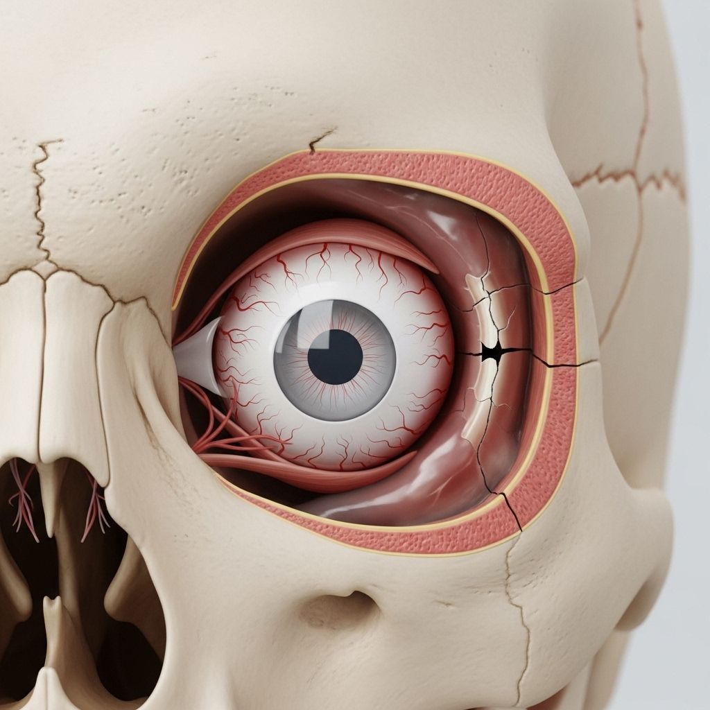
Right Orbital Fracture: Causes, Symptoms, Diagnosis, and Recovery
Understand right orbital fractures—their symptoms, treatment options, and what to expect during recovery for optimal eye health and healing.

Accidents involving facial trauma can result in serious injuries around the eyes, with one such outcome being a right orbital fracture. This injury, involving bones surrounding the right eye socket, requires immediate attention to avoid vision loss, infection, or persistent facial deformity. This guide offers an in-depth exploration of right orbital fractures, covering causes, symptoms, diagnosis, treatment, and recovery, empowering you to make informed decisions if you or someone you know experiences this injury.
What Is a Right Orbital Fracture?
An orbital fracture is a break in one or more bones that form the eye socket (the orbit). When this occurs on the right side, it’s referred to as a right orbital fracture. The orbit is a complex structure made up of several bones: the maxilla, zygomatic, frontal, sphenoid, and ethmoid. Because these bones are thin and delicate in some areas, they are vulnerable to breaks, especially due to blunt trauma.
Causes of Right Orbital Fractures
The most frequent cause of a right orbital fracture is direct trauma to the face. This can happen from:
- Automobile accidents
- Sports injuries (e.g., being hit by a ball or elbow)
- Falls
- Physical assaults
- Workplace or home accidents involving blunt force
The severity of the fracture depends on the force of the trauma and the angle at which it strikes the face.
Types of Orbital Fractures
Right orbital fractures can be classified into several types, depending on which bones are affected:
- Orbital rim fractures: Affect the thick outer edge of the orbit; generally occur due to a strong impact.
- Blowout fractures: Happen when the thin bones at the bottom or inner side of the orbit break, often trapping muscles or tissues.
- Orbital floor fractures: A subtype of blowout fracture involving the floor of the orbit, potentially impacting the maxillary sinus below.
- Orbital roof fractures: Less common, typically seen in children or from high-impact trauma.
Symptoms of a Right Orbital Fracture
Symptoms often depend on the type and severity of the fracture, but common signs include:
- Double, weakened, or blurry vision
- Bruising and swelling around the right eye
- Pain in the cheek, particularly with mouth movement
- Flatness or loss of contour in the cheek area
- Sunken (enophthalmos) or bulging (proptosis) appearance of the right eye
- Numbness or tingling on the right side of the face
- Redness or blood in the white of the eye (subconjunctival hemorrhage)
- Difficulty controlling eye movements (possible muscle entrapment)
Depending on which structures are involved, additional symptoms may include:
- Bleeding from the nose or into the sinus
- Facial asymmetry
- Paresthesia (abnormal skin sensations) in the cheek and upper lip
- Reduced eye movement, sometimes leading to a stuck or fixed gaze
How Is a Right Orbital Fracture Diagnosed?
Diagnosing a right orbital fracture typically involves the following steps:
- Physical examination: A doctor checks for bruising, eye movement problems, bony deformities, or swelling.
- Assessment of visual acuity: Ensures the eye’s vision hasn’t been lost or severely affected.
- Palpation: Feeling around the bone structure to detect irregularities.
- Imaging: A CT scan is the gold standard, clearly showing fractures and muscle/tissue herniation. Sometimes X-rays are used, but they’re less sensitive.
- Testing facial sensation: Checks for nerve involvement in the injury.
- Evaluating for associated injuries: Important in cases of major trauma where multiple facial bones may be involved.
Prompt diagnosis is crucial—missed orbital injuries can lead to complications such as persistent double vision, chronic pain, or cosmetic deformities.
Treatment Options for Right Orbital Fracture
Treatment depends on the severity, location, and type of fracture, as well as presence of symptoms like vision changes or muscle entrapment.
Conservative, Nonsurgical Management
Many minor right orbital fractures are treated without surgery. Common conservative measures include:
- Ice and cold compresses: Reduces swelling and helps manage pain, especially in the first 48 hours.
- Rest: Limits further injury and promotes healing, especially avoiding contact sports or strenuous activities.
- Antibiotics: Prescribed if the fracture involves the sinuses or there is a risk of infection entering through the nose or oral cavity.
- Decongestants: Used to decrease swelling in the sinus and orbital area and make breathing easier—these may be oral or nasal.
- Pain management: Over-the-counter or prescribed pain medications can relieve discomfort.
Precautions During Recovery
- Avoid nose blowing: Blowing your nose can force air or bacteria from the sinuses into the orbit, risking infection or worsening swelling.
- Monitor for worsening symptoms: Such as vision loss, increased pain, or fever—which could indicate complications.
- Follow-up care: Periodic check-ins may be needed to ensure healing is proceeding as expected.
Surgical Treatment
Surgery is considered under specific circumstances, such as:
- Significant muscle entrapment causing eye movement limitation
- Persistent double vision (diplopia) after swelling subsides
- Large fractures with a high risk of sunken eye (enophthalmos)
- Displacement of bone fragments threatening eye function
- Prolapse of orbital contents (tissues or fat herniating into the sinus)
Surgical options typically include:
- Repositioning herniated tissue
- Reconstructing the orbital wall using implants or plates
- Repairing damaged muscles and removing bone fragments
Most surgeries are performed via minimally invasive (transconjunctival or transmaxillary) approaches to reduce scarring. Antibiotics and sometimes corticosteroids may be used post-surgery to prevent infection and reduce swelling. Endoscopic techniques are also increasingly available for targeted repairs.
When Is Surgery Recommended?
A surgeon will consider the following indications for surgical intervention:
- Entrapment of ocular muscles with impaired movement
- Disruption of more than 50% of the orbital floor
- Progressive visual loss due to pressure or entrapment (retrobulbar hematoma)
- Significant cosmetic deformity or loss of orbital volume
- Persistent symptoms not resolving with conservative management
| Factor | Conservative Therapy | Surgery Recommended |
|---|---|---|
| Minor fracture, no muscle herniation | Yes | No |
| Major fracture, muscle entrapment | No | Yes |
| Significant double vision | No | Yes |
| Mild swelling, no vision changes | Yes | No |
Recovery and Outlook
The recovery process for a right orbital fracture depends on the treatment plan:
- Minor fractures: Recovery time is typically a few weeks, with most swelling and bruising resolving within 2 weeks when managed with ice, rest, and medications.
- Surgical cases: Recovery may take several weeks to months. Activities are limited for 4–6 weeks and strenuous exercise should be avoided until cleared by a doctor.
- Potential complications: Chronic double vision, persistent numbness, infection, or cosmetic irregularities may occur and need further management.
Full return to normal activities is generally possible after a proper healing period and with medical clearance. Eye protection is advised during physical activities in the future.
Possible Complications
- Chronic or permanent double vision (diplopia)
- Loss of facial sensation (due to nerve damage)
- Persistent sunken eye appearance (enophthalmos)
- Infection spreading into the sinus or orbital tissues
- Cosmetic deformities requiring further correction
- Traumatic cataract or retinal injury from severe cases
Preventing Orbital Fractures
While accidents may not always be avoidable, certain measures can help reduce the risk of orbital fractures:
- Wearing helmets or face shields during high-risk sports or activities
- Using seat belts and airbags in motor vehicles
- Avoiding physical altercations
- Ensuring a safe home and work environment to minimize falls and sharp objects exposure
Frequently Asked Questions (FAQs)
What is a right orbital fracture?
A right orbital fracture is a break in one or more of the bones that make up the socket surrounding the right eye. It most commonly occurs from direct trauma to the face, such as in car accidents, sports injuries, falls, or assaults.
What symptoms should I look for with a suspected right orbital fracture?
Key symptoms include swelling and bruising around the right eye, double or blurred vision, difficulty moving the eye, pain, sunken or bulging eye, numbness in the cheek/upper lip, and possible blood in the white of the eye.
Will all right orbital fractures require surgery?
No, many minor right orbital fractures heal with conservative measures like ice, rest, antibiotics (if needed), and pain control. Surgery is only necessary if there is significant muscle entrapment, double vision, eye displacement, or a large defect in the orbital wall.
How long does recovery from a right orbital fracture take?
Recovery time can range from a few weeks to several months depending on the severity. Minor fractures may resolve within 2-4 weeks, while severe or surgical cases could require longer healing and follow-up.
Can a right orbital fracture affect my vision permanently?
If treated promptly, most vision problems resolve, but complications such as chronic double vision or persistent numbness can occur in some cases, especially with severe injuries or delayed treatment.
Should I avoid any activities after a right orbital fracture?
Yes. Avoid contact sports, strenuous exercise, and nose blowing during the healing phase. Always consult a healthcare professional before resuming activities to ensure proper healing and prevent complications.
When should I see a doctor for a possible orbital fracture?
Seek medical attention after any facial trauma, especially if you experience visual disturbances, swelling, bruising, difficulty moving your eye, or pain. Sudden vision loss requires emergency assessment.
Key Takeaways
- A right orbital fracture involves the bones around your right eye and usually results from direct blow/trauma to the face.
- Mild fractures often heal with conservative care; severe fractures may require surgical repair, especially to restore eye movement or facial structure.
- Symptoms include swelling, pain, vision changes, and altered eye position. Prompt diagnosis and treatment are crucial.
- With proper management, most people recover well, though complications can arise without timely care.
References and Further Reading
- Basta NM, et al. (2021). Refining indications for orbital floor fracture reconstruction: A risk-stratification tool predicting symptom development and need for surgery.
- Boyd K. (2017). What is an orbital fracture?
- Seen S, et al. (2020). Orbital implants in orbital fracture reconstruction: A ten-year series.
References
- https://www.healthline.com/health/eye-health/right-orbital-fracture
- https://www.templehealth.org/services/conditions/orbital-fractures
- https://www.ncbi.nlm.nih.gov/books/NBK534825/
- https://www.mayoclinic.org/medical-professionals/trauma/news/a-blow-to-the-eye-ocular-and-orbital-trauma/mac-20429287
- https://eyewiki.org/Orbital_Roof_fractures
- https://www.uhsussex.nhs.uk/resources/orbital-fractures-2/
- https://www.childrenshospital.org/conditions/eye-socket-fracture
- https://www.cedars-sinai.org/health-library/diseases-and-conditions—pediatrics/f/fractures-of-the-orbit.html
Read full bio of medha deb


