Pupillary Disorders: Understanding Anisocoria and Related Conditions
Explore the causes, diagnosis, and treatment of pupillary disorders, including anisocoria, for improved eye and neurological health.

Pupillary disorders encompass a range of conditions that affect the size, shape, and reactivity of the pupils. Of these disorders, anisocoria—the presence of unequal pupil sizes—is among the most commonly recognized. While minor variations in pupil size are seen in healthy individuals, more pronounced or persistent inequalities can be a marker of underlying neurological, ocular, or systemic disease. This article provides a detailed examination of pupillary disorders, their underlying causes, clinical implications, and recommended management strategies.
What Are Pupillary Disorders?
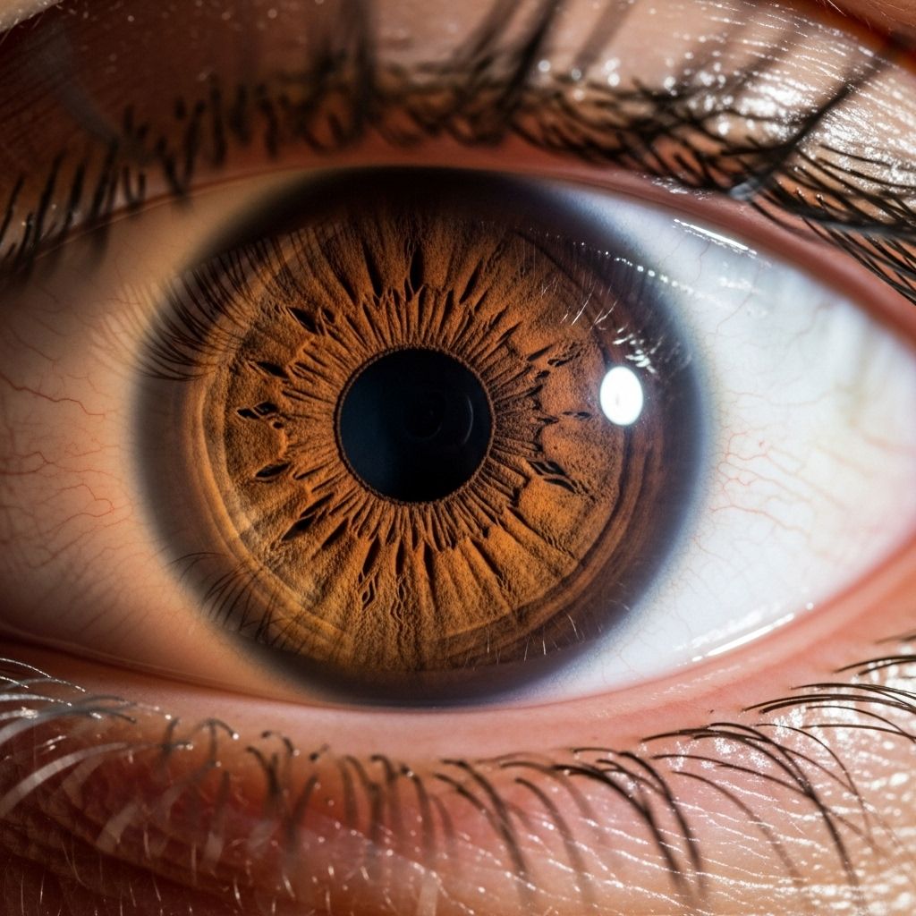
The pupil is the dark, circular opening in the center of the iris that controls the amount of light entering the eye. Under normal circumstances, both pupils should be round and of equal size, responding symmetrically to changes in light intensity. Pupillary disorders arise when these basic features are disrupted, either due to structural abnormalities, muscular dysfunction, or nerve damage.
- Anisocoria: Unequal pupil sizes.
- Pupillary nonreactivity: Pupils fail to respond to light or accommodation.
- Pupillary shape irregularities: The normal round outline of the pupil is lost.
- Pupillary tonic or fixed responses: Pupils dilate or constrict and remain fixed, not responding to further stimuli.
Each of these manifestations can result from different causes and may require tailored diagnostic and treatment approaches.
Defining Anisocoria
Anisocoria is the medical term for unequal pupil size. Typically, differences of less than 1 mm are considered within the range of normal variation, known as physiologic anisocoria. About 1 in 5 people may have such benign, harmless pupil size differences at some time in their life. However, when the difference is more significant or associated with other symptoms, it may reflect an underlying disease or injury affecting the eye or nervous system.
- Physiologic anisocoria is common, mild, and not associated with other symptoms.
- Pathologic anisocoria may be associated with vision changes, pain, eyelid droop, or neurological deficits.
How Do Pupils Normally Function?
The pupils naturally expand (dilate) in low light and contract (constrict) in bright light. This is controlled by two sets of muscles:
- The sphincter pupillae (controlled by the parasympathetic nervous system) constricts the pupil in response to light.
- The dilator pupillae (controlled by the sympathetic nervous system) dilates the pupil in darkness or during excitement.
If the nerves or muscles regulating the pupils are damaged or malfunctioning, pupillary responses become abnormal, often resulting in disorders like anisocoria.
Common Causes of Anisocoria
The causes of anisocoria can range from harmless variants to urgent, life-threatening conditions. Below is an overview of typical etiologies:
| Cause | Key Characteristics |
|---|---|
| Physiologic anisocoria | Very common, usually less than 1 mm pupil difference, no other symptoms. |
| Horner syndrome | Pupil on affected side is smaller, accompanied by drooping eyelid (ptosis), decreased sweating (anhidrosis), delay in pupil dilation. May indicate nerve injury or tumor. |
| Third cranial nerve palsy | Enlarged pupil with droopy eyelid. Can signal compression from aneurysm or tumor. |
| Eye trauma or surgery | May affect iris muscles or nerves. |
| Medication effects | Eye drops, inhaled drugs, or accidental contact with certain medications. |
| Infections or inflammatory conditions | Uveitis, herpes, or other infections affecting the eye. |
| Congenital iris defects | Present from birth; may be associated with other eye or systemic abnormalities. |
While many cases are benign, especially when the difference is stable over time, new or rapidly changing anisocoria may indicate a medical emergency, such as brain aneurysm, stroke, or acute nerve injury.
Symptoms and When to Seek Care
Most people discover anisocoria when they or someone else notices a difference in their pupil sizes. Additional symptoms may suggest a more serious cause and require prompt evaluation. Call for urgent medical care if anisocoria is accompanied by:
- Sudden or severe headache
- Eye pain or discomfort
- Blurred or double vision
- Droopy eyelid
- Weakness, numbness, difficulty speaking or walking
- Loss of consciousness or confusion
Anisocoria alone does not cause blindness, but its underlying causes—especially if involving major nerves or the brain—can threaten sight and life.
Diagnosing Pupillary Disorders
A thorough diagnosis of pupillary abnormalities involves several steps:
- History: Questions are asked about the duration and onset of symptoms, associated pain, trauma, birth history, medication exposures, and any other neurological symptoms.
- Physical Exam: The doctor will evaluate pupil size and shape in both light and dark, check their reactivity to light and accommodation, assess eyelids, ocular structures, and sometimes compare to prior photos or identification cards.
- Additional tests: Depending on findings, tests may include slit-lamp examination, pupillometry, blood tests, imaging (CT, MRI), and specific pharmacologic tests (e.g., using eye drops to test nerve function).
The goal of the evaluation is to determine if the anisocoria is physiologic (benign) or pathologic (possibly dangerous), and if so, to identify its underlying cause.
Special Considerations in Children and Congenital Anisocoria
Anisocoria present from birth (congenital anisocoria) is usually benign, especially when stable and not associated with other abnormalities. However, some congenital causes may be linked to genetic or developmental problems affecting the iris or nervous system. A detailed eye examination and review of family or birth history are key in these cases.
Specific Pupillary Disorders Related to Anisocoria
- Adie’s Tonic Pupil: One pupil is larger and reacts slowly to light but better to focusing on near objects. Often due to nerve damage after a viral illness or trauma.
- Argyll Robertson Pupil: Small, irregular pupils that react poorly to light but constrict when focusing on near objects. Classically associated with late-stage syphilis.
- Horner Syndrome: Characterized by ptosis (drooping eyelid), miosis (small pupil), and anhidrosis (lack of sweating), usually due to disruption of the sympathetic nerves.
- Third Nerve Palsy: May present with a large, unreactive pupil, ptosis, and impaired eye movement—often a neurologic emergency.
- Pharmacologic Anisocoria: Occurs with accidental or intentional use of certain medications or substances. Will often resolve when the drug wears off, though specialist input is sometimes needed.
How Is Anisocoria Evaluated?
Evaluation follows a systematic approach. The steps commonly include:
- Comparing size in light and dark environments to determine which pupil is behaving abnormally.
- Assessing the speed and degree of reactivity to light and focusing on near objects.
- Evaluating presence of other eye or neurological abnormalities such as eyelid droop, double vision, or facial sweating differences.
- Reviewing photographs or IDs for evidence of chronicity.
- Ordering further diagnostic tests based on clinical suspicion (e.g., imaging for suspected brain involvement).
Treatment of Pupillary Disorders
Treatment for anisocoria depends entirely on its underlying cause.
- Physiologic anisocoria requires no treatment at all.
- Pathologic anisocoria requires treatment tailored to the underlying condition:
- Horner syndrome: Identifying and treating the underlying cause (e.g., tumor, vascular lesion).
- Third nerve palsy: Treating aneurysms, tumors, or infections as indicated; may require urgent intervention.
- Infections or inflammatory diseases: Managing infections or inflammation with medications.
- Medication-induced: Discontinuing the causative drug if possible.
Most people with benign, physiologic anisocoria require no interventions, and patient education on the benign nature of their condition is important to prevent unnecessary worry.
Prognosis and Complications
The prognosis is excellent for benign cases. However, when anisocoria results from neurological or vascular causes, outcomes depend on timely diagnosis and effective management of the underlying condition. Potential complications include:
- Persistent vision problems.
- Functional and cosmetic concerns.
- Severe complications if due to stroke, aneurysm, or tumors (including risk to life).
Frequently Asked Questions (FAQs)
Q: Can anisocoria make you blind?
A: No, anisocoria itself cannot cause blindness. However, the underlying conditions responsible for new or worsening anisocoria may threaten vision if not addressed promptly.
Q: Is a small difference in pupil size normal?
A: Yes, small and stable differences (usually less than 1 mm) between pupil sizes are common and considered physiologic in up to 20% of people.
Q: When should someone seek immediate medical care for anisocoria?
A: Seek immediate care if unequal pupils arise suddenly and are accompanied by severe headache, vision loss, double vision, eyelid droop, confusion, or other neurological symptoms.
Q: Can medications cause anisocoria?
A: Yes. Certain eye drops or accidental exposure to agents like scopolamine, pilocarpine, or inhaled medications can cause transient pupil size differences. Usually, this resolves after stopping the medication.
Q: What tests are typically done to diagnose the cause of anisocoria?
A: Tests may include a detailed eye examination, pupillary response assessments, review of old photos, neurological assessment, imaging (MRI, CT), and possibly pharmacologic pupil testing.
Prevention and Lifestyle Considerations
While most cases of anisocoria cannot be prevented, minimizing risk factors such as avoiding eye injuries and appropriately using prescription or over-the-counter medications can help. Being aware of symptoms and rapidly seeking care when new or severe anisocoria occurs is vital.
- Use medications only as prescribed, especially those intended for the eyes.
- Wear eye protection during sports or hazardous activities.
- Report any sudden change in vision, pupil size, or eye pain to your healthcare provider.
Key Points
- Physiologic anisocoria is common and benign; differences greater than 1 mm or with symptoms require evaluation.
- Serious disorders should be considered in cases with Horner syndrome or third nerve palsy.
- Evaluation in both light and dark, and comparison to old photos, can help identify abnormal pupils.
- Treatment focuses on the underlying disease, not the anisocoria itself.
Resources and Support
- Consult with an ophthalmologist or neuro-ophthalmologist if anisocoria or related symptoms are present.
- The North American Neuro-Ophthalmology Society and American Academy of Ophthalmology provide further information and support resources.
- Local emergency services should be utilized if anisocoria is accompanied by acute neurological changes.
References
- https://www.merckmanuals.com/professional/eye-disorders/symptoms-of-ophthalmic-disorders/anisocoria
- https://www.nanosweb.org/anisocoria/
- https://my.clevelandclinic.org/health/diseases/22422-anisocoria
- https://med.stanford.edu/stanfordmedicine25/the25/pupillary.html
- https://pubmed.ncbi.nlm.nih.gov/40179407/
- https://pubmed.ncbi.nlm.nih.gov/21601076/
Read full bio of Sneha Tete


