Positron Emission Tomography (PET): Diagnostic Imaging and Clinical Impact
A comprehensive guide to PET scans exploring their purpose, procedure, safety, and evolving clinical applications in modern medicine.

Positron Emission Tomography (PET) is an advanced functional imaging technology that plays an essential role in diagnosing, staging, and monitoring a range of diseases. By providing vivid, real-time images of the inside of the body, particularly cellular activity and metabolism, PET scans have become an indispensable tool in oncology, neurology, cardiology, and emerging fields. This article delivers a detailed exploration of PET technology, its uses, preparation, procedure, risks, and frequently asked questions, reflecting current best practices in modern medicine.
What is a PET Scan?
A Positron Emission Tomography (PET) scan is a nuclear medicine imaging technique used to observe metabolic processes in the body. It achieves this by using a small amount of radioactive material (referred to as a radiotracer), which is introduced into the body either by injection, inhalation, or orally. Special cameras then detect the gamma rays emitted from the tracer to produce detailed images of tissue function and cell activity. These images often highlight areas of abnormal metabolism, which is crucial for identifying conditions like cancer, heart disease, and brain disorders.
Why Might I Need a PET Scan?
PET scans are versatile diagnostic tools that support medical decision-making in a variety of clinical settings. Reasons a provider may recommend a PET scan include:
- Cancer: Detecting, staging, and monitoring the progression or recurrence of various cancers, including lung, breast, colorectal, and lymphoma.
- Neurology: Assessing brain disorders such as Alzheimer’s disease, epilepsy, and Parkinson’s disease by visualizing chemical activity in brain cells.
- Cardiology: Evaluating blood flow, detecting areas of reduced perfusion, and assessing tissue viability in ischemic heart disease.
- Infectious or Inflammatory Diseases: Identifying sites of active inflammation or chronic infection, particularly in diseases that affect multiple organs.
Types of PET Scans
While the traditional PET scan focuses on metabolic activity, technological advances have led to various PET-based imaging techniques for targeted diagnosis. The main types include:
- Standard PET: Uses tracers such as fluorodeoxyglucose (FDG) to assess glucose metabolism.
- PET/CT: Combines PET and computed tomography for both metabolic and anatomical precision, allowing for accurate localization of abnormalities.
- PET/MRI: Merges PET’s metabolic insights with magnetic resonance imaging’s soft-tissue detail, increasingly used in neurology and pediatric oncology.
How Does a PET Scan Work?
PET scans rely on the principles of nuclear medicine and radioactive decay. The basic steps are:
- Tracer Administration: A radioactive tracer (often FDG) that mimics natural substances like glucose is administered to the patient.
- Cellular Uptake: The tracer travels throughout the body and accumulates in areas of high metabolic activity (e.g., tumors, inflamed tissues, or active brain regions).
- Emission Detection: As the tracer decays, it emits positrons that interact with electrons, resulting in the release of gamma rays.
- Image Reconstruction: Specialized cameras detect the gamma rays, and computer algorithms reconstruct detailed images showing the biodistribution of the tracer.
Clinical Applications of PET Scans
| Field | Primary Uses | Example Conditions |
|---|---|---|
| Oncology |
| Lymphoma, lung, breast, colorectal cancers |
| Neurology |
| Gliomas, Alzheimer’s disease, seizure disorders |
| Cardiology |
| Coronary artery disease, infarctions |
| Other Fields |
| Sarcoidosis, vasculitis, chronic infections |
Preparing for a PET Scan
Preparation for a PET scan maximizes the accuracy and safety of the procedure. Some general guidelines include:
- Diet: Patients may be advised to avoid eating for several hours (often four to six) before the scan, as high blood sugar levels can interfere with tracer uptake.
- Medication: It is important to inform your medical team about all medications. Certain medicines may need to be paused or adjusted before the test.
- Health History: Notify your healthcare provider if you are pregnant, breastfeeding, diabetic, or have allergies to contrast materials or iodine.
- Comfort: Wear comfortable clothing and remove any metal objects, such as jewelry, which can interfere with imaging.
What Happens During the PET Scan Procedure?
The PET scan process typically follows these steps:
- Arrival and Preparation: Upon arriving at the imaging center, you may change into a gown and provide a full medical history.
- Tracer Administration: The radiotracer will be administered, usually by injection into a vein but sometimes via inhalation or orally.
- Absorption Period: After the tracer is given, you’ll rest quietly for 30 to 90 minutes to allow your body to absorb and distribute it. Movement and talking should be minimized to enhance image clarity.
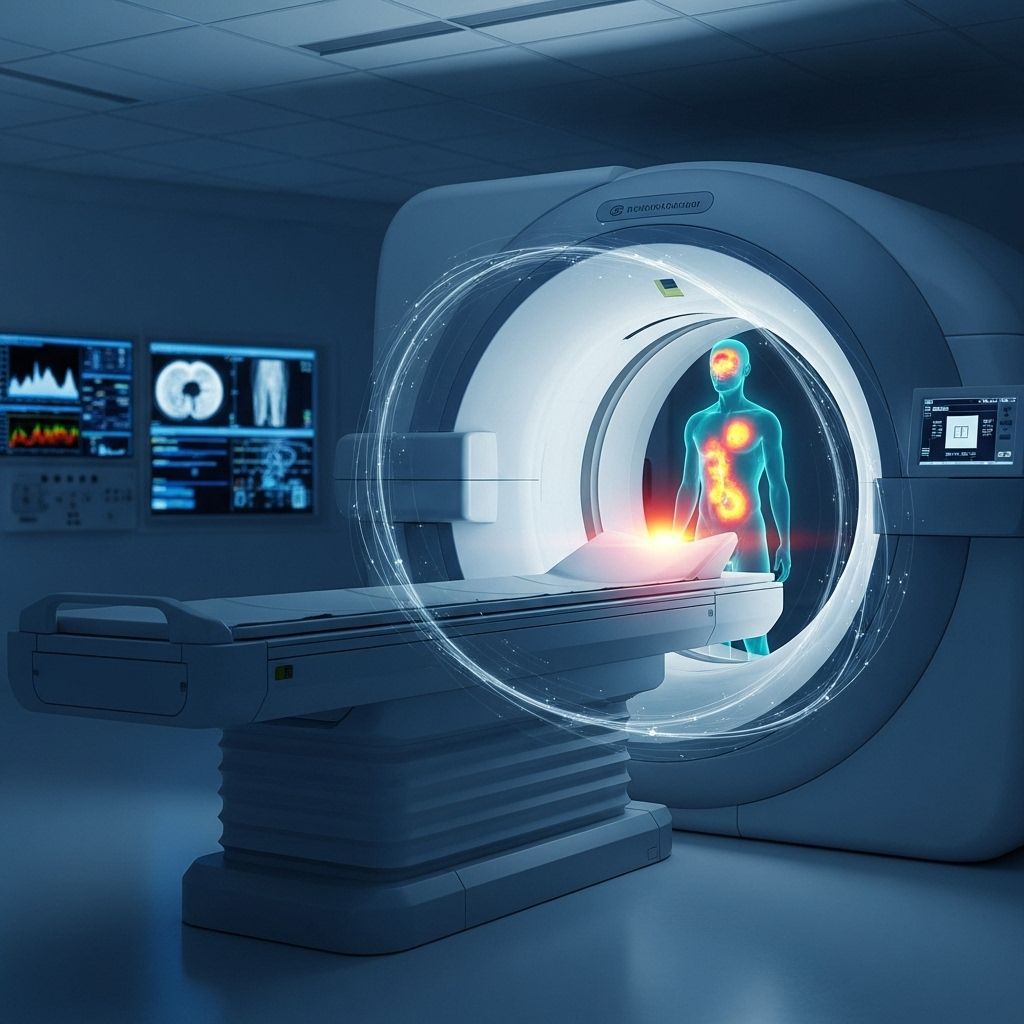
- Imaging: You will lie still on a padded table that slides slowly into the PET scanner (a large, ring-shaped instrument). The scan usually takes 30 to 60 minutes, during which you must remain as still as possible for optimal image acquisition.
- Completion: When imaging is complete, you’re typically free to leave unless further observation is needed.
After the PET Scan: What to Expect
Most individuals can return to normal activities shortly after the scan. Important post-scan considerations include:
- Hydration: Drink plenty of fluids to accelerate the elimination of the tracer from your system.
- Interacting With Others: To be extra cautious, avoid close contact with pregnant women or small children for several hours following the test.
- Results: A radiologist will analyze the images and send a detailed report to your referring physician, who will discuss the findings with you.
Safety and Risks of PET Scans
PET scans are generally considered safe and are non-invasive. However, as with any medical procedure that involves radiation or injection, there are specific risks:
- Radiation Exposure: The amount of radiation used is relatively low and is quickly eliminated from the body. The benefits typically outweigh the minimal risks.
- Allergic Reactions: Rarely, patients may experience an allergic response to the radioactive tracer.
- Discomfort or Bruising: Some discomfort may occur at the injection site.
- Pregnancy and Breastfeeding: Special precautions are required. Women who are or may be pregnant, or who are breastfeeding, should inform their provider.
- Special Populations: Patients with kidney problems, diabetes, or allergies should discuss their conditions with the medical team in advance.
Advancements in PET Imaging Technology
Recent innovations have enhanced PET technology’s utility and safety:
- Total-Body PET/CT: Provides whole-body imaging in a single scan with reduced radiation and shorter acquisition times, making it more accessible for pediatric or critically ill patients.
- Hybrid Imaging: PET/CT and PET/MRI combine metabolic and structural data, improving diagnostic accuracy and guiding precision therapies for complex diseases.
- New Radiotracers: Ongoing research is developing novel tracers for improved specificity in cancer, neurology, cardiology, and infection imaging, broadening the scope and precision of PET scans.
Limitations of PET Scans
Despite their advantages, PET scans have certain limitations:
- Resolution: PET images typically have lower spatial resolution than CT or MRI.
- Cost and Availability: PET scans are more expensive and not as widely available as some other imaging techniques.
- Preparation Sensitivity: Improper preparation can affect accuracy, especially in diabetes or recent exercise, which alters glucose metabolism.
- False Positives/Negatives: Non-cancerous inflammation or infection may mimic malignancy, while certain slow-growing tumors may not show increased uptake.
Frequently Asked Questions (FAQs)
Q: How do PET scans differ from CT or MRI?
A: CT and MRI provide anatomical images using X-rays or magnetic fields, showing structures and shapes inside the body. PET, in contrast, reveals functional processes and cellular metabolism. PET/CT and PET/MRI combine these methods for comprehensive insights.
Q: Are PET scans painful?
A: The scan itself does not cause pain. Patients may feel a brief pinch from the tracer injection but usually experience no discomfort during imaging.
Q: How long does it take to get PET scan results?
A: The images are typically interpreted by a radiologist within 24–48 hours. The ordering provider will share results and discuss next steps with you soon after.
Q: Can I drive home after a PET scan?
A: Yes, most patients can drive themselves home unless sedation was used or they feel unwell after the procedure.
Q: Are there alternatives to PET scans?
A: Alternatives include CT, MRI, or SPECT scans. The best test depends on the clinical question, organ system, and condition being investigated. Your provider can advise on the most appropriate choice for your needs.
Key Takeaways
- PET scans provide critical functional information about tissues and organs, complementing anatomical imaging.
- They are vital in cancer diagnosis, neurological disease assessment, and heart disease evaluation.
- Preparation is important for obtaining accurate results—follow your healthcare team’s instructions carefully.
- Risks are minimal, but discuss any health conditions or concerns with your provider before the test.
Additional Resources
- Society of Nuclear Medicine and Molecular Imaging (SNMMI)
- American College of Radiology (ACR): Imaging Tests
- National Cancer Institute: PET Scans
References
- https://www.ganeshdiagnostic.com/blog/clinical-applications-of-pet-scans-a-review-of-common-uses
- https://pmc.ncbi.nlm.nih.gov/articles/PMC4921358/
- https://ajronline.org/doi/full/10.2214/ajr.19.22705
- https://pmc.ncbi.nlm.nih.gov/articles/PMC11014840/
- https://www.mayoclinic.org/tests-procedures/pet-scan/about/pac-20385078
- https://www.radiologyinfo.org/en/info/pet
- https://acsjournals.onlinelibrary.wiley.com/doi/full/10.1002/cncr.28860
- https://health.ucdavis.edu/radiology/myexam/PET/Equipment/explorer.html
Read full bio of Sneha Tete


