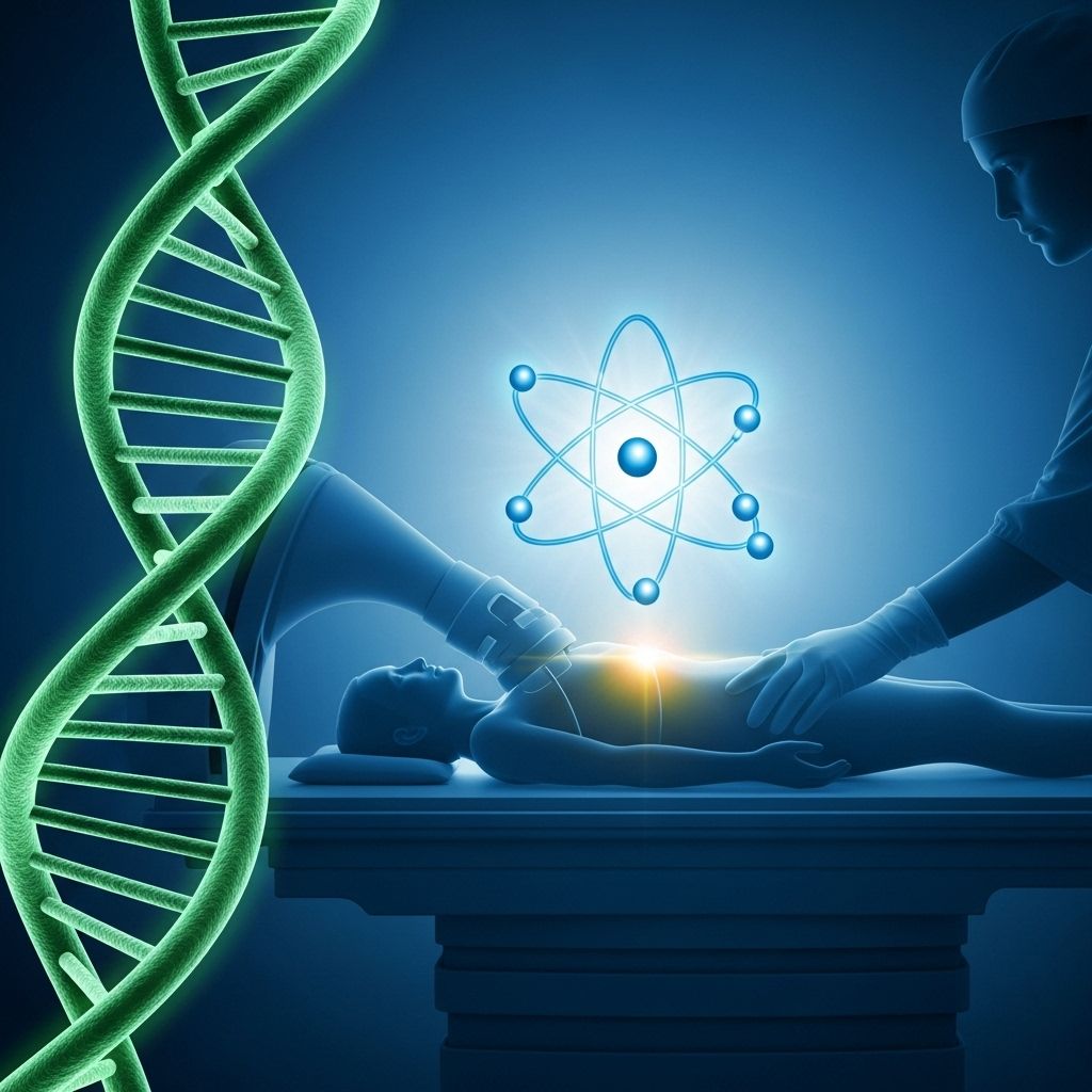
Nuclear Medicine: Principles, Procedures, and Patient Care
Explore how nuclear medicine uses radiopharmaceuticals for diagnosing and treating disease, offering unique insights into organ function and advanced therapies.

Nuclear medicine is a specialized area of medical imaging that uses small amounts of radioactive materials, known as radiopharmaceuticals, to diagnose and treat a wide variety of diseases. Unlike conventional radiology, which reveals anatomical structures, nuclear medicine uniquely assesses the function and physiology of organs and tissues.
This article provides a detailed overview of nuclear medicine, including its techniques, common uses, patient experience, safety considerations, and frequently asked questions.
What Is Nuclear Medicine?
Nuclear medicine is a branch of radiology that introduces radiopharmaceuticals into the body to visualize, diagnose, and sometimes treat disease. These substances can be injected, swallowed, or inhaled. As the radioactive drug accumulates in specific organs or tissues, it emits radiation that is captured by special cameras to create images or track organ function.
Unlike traditional imaging that highlights only the structure of organs, nuclear medicine assesses how organs are working. This capability allows clinicians to identify dysfunction at an early stage, often before structural changes become apparent on CT or MRI scans.
Key Features of Nuclear Medicine
- Functional Imaging: Reveals how organs and tissues are functioning, not just their appearance.
- Early Disease Detection: Can reveal disease in early or even preclinical stages.
- Personalized Therapy: Certain radiopharmaceuticals are used therapeutically, targeting tumors or abnormal tissues.
- Low Radiation Dose: Utilizes very small amounts of radioactive material, generally resulting in minimal risk.
How Does Nuclear Medicine Work?
During a nuclear medicine procedure, a radiopharmaceutical is administered to the patient. The type of drug used depends on the organ or physiological process to be examined. These compounds are designed to localize in specific tissues, such as the heart, bones, thyroid, or brain.
Once in the body, these substances emit gamma rays or positrons, which are detected by external cameras and processed to produce detailed images or quantitative information about organ function.
Types of Imaging Techniques
- Gamma Camera Imaging (Scintigraphy): Detects gamma rays emitted by the radiopharmaceutical, producing two-dimensional images.
- Single Photon Emission Computed Tomography (SPECT): Produces three-dimensional images by capturing gamma rays from multiple angles.
- Positron Emission Tomography (PET): Uses positron-emitting radiopharmaceuticals for highly sensitive 3D imaging, often combined with CT (PET/CT) for enhanced anatomical localization.
Common Uses for Nuclear Medicine
Nuclear medicine offers distinct advantages in evaluating a broad range of organ systems. Below are some of its most frequent applications:
- Heart:
- Assessing coronary artery disease (myocardial perfusion scans).
- Evaluating heart function (ventricular ejection fraction studies).
To better understand the purpose and process of a Heart Perfusion Scan, it’s essential to explore our detailed explanation. This scan is crucial for assessing heart health and can provide peace of mind, especially for those with cardiovascular concerns. - Bones:
- Detecting bone infections, fractures, or the spread of cancer to bones (bone scans).
- Thyroid Gland:
- Diagnosing thyroid function, overactivity (hyperthyroidism), or identifying thyroid cancer.
- Kidneys and Bladder:
- Evaluating kidney function or urinary tract obstruction.
- Lungs:
- Detecting blood clots (pulmonary embolism) using ventilation/perfusion (V/Q) scans.
- Cancer:
- Detecting, staging, and monitoring treatment response in various types of cancer using PET scans.
- Treating some cancers, such as thyroid cancer, with targeted radiotherapy.
How Should I Prepare for The Procedure?
Preparation for a nuclear medicine exam varies depending on the specific test. Your healthcare provider will give you detailed instructions tailored to your test and medical history. Common preparation steps include:
- Fasting (not eating or drinking for a certain time) before the procedure, especially for PET scans or gallbladder studies.
- Stopping some medications that may interfere with test accuracy.
- Wearing comfortable clothing; you may be asked to change into a hospital gown.
- Informing your healthcare team if you are pregnant, breastfeeding, or have allergies to any medications.
What Happens During a Nuclear Medicine Procedure?
While each test has unique steps, the general process in nuclear medicine exams follows this sequence:
- Radiopharmaceutical Administration: The radioisotope is injected into a vein, swallowed, or inhaled. Which route is used depends on the organ to be studied and the exam type.
- Uptake Period: There may be a waiting period ranging from a few minutes to several hours to allow the radiopharmaceutical to accumulate in the target organ.
- Imaging Session: You will be positioned on a specialized table, and a gamma camera or PET scanner will capture images while you remain still.
- Test Duration: Most nuclear medicine scans last between 30 minutes and several hours. Some procedures may require images at multiple time points.
What Happens After a Nuclear Medicine Procedure?
After the exam:
- Most patients can resume normal activities immediately after the procedure.
- You will be encouraged to drink fluids to help flush any remaining radioactive material from your body.
- The small amount of radioactivity typically disappears within hours to a few days, depending on the substance used.
- Your imaging results will be interpreted by a nuclear medicine specialist and shared with your referring doctor, who will explain the findings to you and plan next steps.
What Are the Risks and Potential Complications?
Nuclear medicine procedures involve exposure to a low dose of radiation. The risks are generally minimal, and the benefits of accurate diagnosis or treatment almost always outweigh potential harms.
- Allergic reactions or side effects from radiopharmaceuticals are extremely rare.
- Most nuclear medicine scans deliver a dose of radiation similar to or less than a routine diagnostic X-ray.
- Pediatric patients and pregnant women should only undergo nuclear medicine examinations when absolutely necessary, as recommended by medical guidelines.
It is crucial to inform your healthcare provider if you are or think you may be pregnant, or if you are breastfeeding, prior to the exam. Precautions can be taken to minimize or avoid exposure in these situations.
What Are the Benefits of Nuclear Medicine?
- Early Disease Detection: Ability to detect changes in organ function often before symptoms develop or structural changes appear on other imaging.
- Functional Assessment: Provides valuable data on physiological processes, helping refine diagnoses and guide therapy.
- Targeted Therapy: Some radiopharmaceuticals deliver therapeutic doses of radiation directly to diseased tissue, sparing healthy tissue.
- Noninvasive: Most procedures do not require incisions, and injections are similar to a routine blood draw.
- Complementary Imaging: Nuclear medicine findings often complement results from CT, MRI, or ultrasound exams, creating a more complete clinical picture.
Who Performs and Interprets Nuclear Medicine Studies?
Nuclear medicine procedures are performed by specially trained technologists and interpreted by physicians who are board-certified in nuclear medicine or radiology. These professionals have expertise in radiation safety, radiopharmaceutical handling, and image interpretation, ensuring high diagnostic quality and patient safety.
Patient Experience: What to Expect
Nuclear medicine tests are generally painless and well-tolerated. You may feel a slight pinch during an injection, but most patients are comfortable throughout the procedure.
For certain studies, you may be asked to remain still for an extended period while images are acquired. Let the technologist know if you need to adjust position or if you experience any discomfort.
Frequently Asked Questions (FAQs)
How safe is nuclear medicine?
Nuclear medicine exams are very safe. The amount of radioactive material used is small, and the risk from radiation exposure is minimal when compared to the benefits of accurate diagnosis and treatment.
Will I be radioactive after the test?
For a short time, you may emit a small amount of radiation, but this quickly diminishes as the material leaves your body. It’s typically safe to be around others, except in rare cases where special precautions will be outlined.
Can children have nuclear medicine scans?
Yes, many childhood illnesses are best diagnosed with nuclear medicine. Pediatric protocols use the lowest possible dose tailored to the child’s weight and needs.
What if I am pregnant or breastfeeding?
Inform your healthcare provider before the test. In some cases, the procedure may be postponed or alternative imaging used; precautions are taken to minimize potential effects on the unborn child or nursing infant.
How should I care for myself after the procedure?
Drink plenty of fluids to help clear the radiopharmaceutical. There are usually no activity restrictions post-procedure, unless advised otherwise by your clinician.
Summary Table: Nuclear Medicine at a Glance
| Aspect | Description |
|---|---|
| Purpose | Diagnosis and treatment using radiopharmaceuticals |
| Imaging Methods | Gamma camera, SPECT, PET, PET/CT |
| Common Uses | Heart, bone, thyroid, lung, kidney, and cancer studies |
| Preparation | May involve fasting, medication changes, or specific instructions |
| Risks | Minimal; small radiation dose similar to X-rays |
| Benefits | Early detection, functional assessment, targeted therapy |
Conclusion
Nuclear medicine is a vital field in modern healthcare, providing unique insight into how the body functions at the molecular level. Its ability to detect disease early, guide therapies, and offer noninvasive treatment options makes it an indispensable diagnostic and therapeutic tool. Advances in radiopharmaceuticals and imaging technology continue to increase the accuracy, safety, and clinical utility of nuclear medicine procedures.
Frequently Asked Questions (FAQs)
Q: How does nuclear medicine differ from X-ray or CT imaging?
A: While X-ray and CT imaging focus on anatomical details, nuclear medicine shows how organs and tissues function, revealing disease processes before changes in structure become visible.
Q: What should I bring to my nuclear medicine appointment?
A: Bring any test orders, insurance information, a list of current medications, and follow all preparation instructions provided by your care team.
Q: Will nuclear medicine procedures affect my daily routine?
A: In most cases, you can return to your normal schedule quickly. It’s generally advised to increase fluids to help eliminate any remaining tracer from your body.
Q: Are there any side effects?
A: Side effects are rare. Most patients do not feel any different after receiving the radiopharmaceutical.
Q: Who provides my results?
A: A nuclear medicine doctor analyzes the images and sends a detailed report to your primary care provider or referring physician, who will discuss the results and next steps with you.
References
- https://somi.jh.edu/programs/nuclear-medicine-technology/
- https://pure.johnshopkins.edu/en/publications/nuclear-medicine-spect-single-photon-emission-computed-tomography
- https://www.youtube.com/watch?v=U0SZHGfP_1A
- https://somi.jh.edu/wp-content/uploads/2024/09/JHSOMI-NM-FAQs-08.2024.pdf
- https://www.hilarispublisher.com/open-access/nuclear-medicine-an-overview-84779.html
- https://coconote.app/notes/30bed7e9-5058-42a4-b6af-6bcb9353af4d
- https://pubmed.ncbi.nlm.nih.gov/26133075/
Read full bio of Sneha Tete


