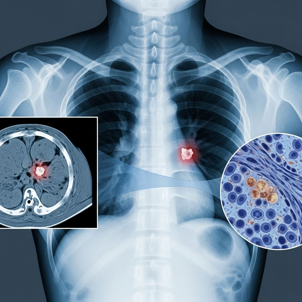
Understanding Non-Small Cell Lung Cancer: Diagnosis Through X-ray and Beyond
Learn how non-small cell lung cancer is identified using X-rays, their limitations, and modern diagnostic advancements.

Non-Small Cell Lung Cancer and X-ray Diagnosis: What You Need to Know
Non-small cell lung cancer (NSCLC) is the most common type of lung cancer, making up about 80-85% of all lung cancer cases worldwide. Like all forms of lung cancer, NSCLC is most treatable when detected early, but obtaining a timely diagnosis can be challenging. One of the primary tools used when symptoms suggest lung cancer is a chest X-ray. While X-rays are widely available and noninvasive, their role in diagnosing NSCLC comes with both strengths and significant limitations. This article explores how NSCLC is detected using X-rays, what you can expect during the process, and how newer imaging technologies are enhancing diagnostic accuracy.
What is Non-Small Cell Lung Cancer?
Non-small cell lung cancer is a term that refers to several types of lung cancer sharing similar behavior and treatment approaches. The three major types of NSCLC are:
- Adenocarcinoma
- Squamous cell carcinoma
- Large cell carcinoma
These cancers tend to grow and spread more slowly compared to small cell lung cancer. Symptoms often do not appear until the disease is at an advanced stage, making early detection crucial for successful treatment.
Understanding Lung X-rays
An X-ray is a quick, safe, and cost-effective imaging technique that uses small doses of radiation to create pictures of the inside of your body. In the context of lung cancer, a chest X-ray produces images of your lungs, heart, airways, and bones.
How a Chest X-ray Works
- X-rays are directed through your chest to a digital detector or film placed behind you.
- Structures that absorb X-rays (like bones or tumors) appear white or light gray.
- Air-filled spaces such as healthy lung tissue appear black.
Chest X-rays are often the first test recommended if you are experiencing symptoms like a persistent cough, chest pain, breathlessness, or unexplained weight loss.
What Non-Small Cell Lung Cancer Looks Like on an X-ray
An area of NSCLC may appear on an X-ray as:
- A distinct, dense white or gray mass, usually outlined against the black of the lung
- Areas of partial or total lung collapse (atelectasis), visible as sections where lung tissue is no longer air-filled
- Obscured or distorted lung outlines, suggesting growths or blockages
- Changes in the silhouette of the heart or diaphragm if the cancer has grown near these areas
- Enlarged lymph nodes near the middle of the chest, sometimes noticeable as abnormal densities
However, X-rays have significant limitations for detecting lung cancer, especially in its early stages. Not all tumors are visible, and benign conditions can look similar to cancer on X-rays.
Limitations of X-rays in Diagnosing NSCLC
While X-rays are helpful for uncovering abnormal areas in the lungs, there are several reasons they may miss or misidentify cancers:
- Small tumors might not show up: Some cancers, especially those less than 1-2 cm, can be missed.
- Overlapping structures: The ribs, heart, and blood vessels can hide or mimic tumors.
- Non-cancerous conditions: Infections, scars, or benign masses sometimes appear similar to cancer.
- No definitive diagnosis: X-rays can suggest something is abnormal, but cannot confirm cancer; further tests are always needed.
Multiple studies show that chest X-rays have low sensitivity for detecting early-stage lung cancer—the accuracy improves for larger or more advanced tumors.
Other Imaging Tests and Diagnostic Steps
If your chest X-ray suggests an abnormality, your healthcare provider will typically order additional tests:
- CT scan (Computed Tomography): Offers more detailed, cross-sectional images and is better at identifying small or hidden tumors.
- PET/CT scan: Combines metabolic and structural imaging to assess the spread (metastasis) and activity of suspicious areas.
- MRI (Magnetic Resonance Imaging): Used in specific cases for detailed imaging, especially evaluating the brain or spinal cord if spread is suspected.
- Biopsy: Involves sampling suspicious tissue to confirm the presence and type of cancer cells. Types include needle biopsy, bronchoscopy, thoracoscopy, or mediastinoscopy.
- Blood tests: Not diagnostic for cancer but help assess overall health and organ function before treatment.
These tests help determine the cancer’s type, exact location, and if it has spread, making them vital for treatment planning.
Comparing X-ray and CT for Lung Cancer
| Feature | Chest X-ray | CT Scan |
|---|---|---|
| Availability | Wide, fast, affordable | Available, higher cost |
| Detail Level | Basic, 2D images | Highly detailed, 3D slices |
| Detection Sensitivity | Lower (may miss small tumors) | Higher (better for early detection) |
| Radiation Dose | Low | Higher than X-ray |
| Definitive for Cancer? | No—cannot confirm cancer | No, but provides better data for further testing |
How the Diagnostic Process Looks for Patients
If your symptoms or history suggest possible lung cancer, here is a typical diagnostic journey you might experience:
- Initial Evaluation: Your healthcare provider will discuss symptoms (like cough, chest pain, weight loss, or fatigue) and any risk factors, such as smoking history or family cancer history.
- Physical Examination: May include listening to your lungs, checking breathing, and sometimes basic lung function tests (spirometry).
- Chest X-ray: The first imaging test in most cases to look for lung abnormalities.
- Follow-up Imaging: If the X-ray suggests something unusual or is inconclusive, a CT scan or other imaging is arranged for greater clarity.
- Specialist Referral: You’ll be referred to a pulmonary or oncology specialist for evaluation and further diagnostic steps.
- Biopsy: To confirm cancer and its specific type.
- Staging: Determining the extent of disease spread with additional imaging or procedures.
Remember: A chest X-ray is the starting point—not the final answer. A normal chest X-ray does not completely exclude lung cancer, especially in the early phase.
What Else Can a Chest X-ray Show?
Besides cancer, a chest X-ray can help reveal:
- Lung infections (pneumonia, abscess)
- Chronic lung diseases (COPD, fibrosis)
- Fluid around the lungs (pleural effusion)
- Heart size and shape anomalies
- Fractures of the ribs or spine
This underscores the importance of specialist follow-up, as many non-cancerous lung diseases can mimic or mask lung cancer on an X-ray.
What Should You Do if an X-ray Shows an Abnormality?
- Do not panic—a suspicious shadow does not always mean cancer. Many benign conditions cause similar findings.
- Follow your healthcare provider’s recommendations for further testing or specialist referral.
- Keep track of new or worsening symptoms and report them promptly.
Frequently Asked Questions (FAQs)
Q: Can a chest X-ray alone diagnose non-small cell lung cancer?
A: No. A chest X-ray can show suspicious lung shadows or masses, but cannot definitively diagnose cancer. Further imaging and a biopsy are always required.
Q: What are the signs of non-small cell lung cancer on an X-ray image?
A: NSCLC may show up as a solid white or gray spot, an area of lung collapse, or enlarged lymph nodes. However, some tumors are too small to be seen or are hidden by other structures.
Q: Are there risks associated with having a chest X-ray?
A: Chest X-rays use a very small amount of radiation, generally considered safe. Risks are minimal but discuss any concerns with your provider, especially if you require repeated imaging.
Q: If my X-ray is normal but I still have symptoms, what should I do?
A: Talk to your healthcare provider about your ongoing symptoms. Further tests, such as a CT scan or referral to a specialist, may be needed as some early lung cancers are not visible on X-ray.
Q: What are the next steps if lung cancer is suspected based on imaging?
A: You will usually have a CT scan next. If abnormalities persist, a biopsy is performed to confirm the presence and type of cancer cells, followed by staging tests to guide treatment.
Key Takeaways
- Chest X-ray is often the first test when lung cancer symptoms are present, but it cannot provide a definitive diagnosis on its own.
- Small tumors are often invisible or unclear on X-rays, so further testing is essential if clinical suspicion remains high.
- Modern imaging—especially CT and PET/CT—have improved early detection and accurate staging of NSCLC.
- Always follow up with your healthcare provider for a complete diagnosis and treatment plan.
When to See a Doctor
If you experience persistent symptoms such as a cough, chest pain, shortness of breath, coughing up blood, or unexplained weight loss, contact your healthcare provider. Early evaluation and testing improve the chance of a curable outcome.
References
- This article is based on current expert consensus and reputable health resources. For personalized advice, always consult your specialized medical provider.
References
- https://pmc.ncbi.nlm.nih.gov/articles/PMC6609262/
- https://my.clevelandclinic.org/health/diseases/4375-lung-cancer
- https://nyulangone.org/conditions/non-small-cell-lung-cancer/diagnosis
- https://www.nhs.uk/conditions/lung-cancer/diagnosis/
- https://www.cancer.org/cancer/types/lung-cancer/detection-diagnosis-staging/detection.html
- https://www.dana-farber.org/cancer-care/types/non-small-cell-lung-cancer/diagnosis
- https://www.mayoclinic.org/diseases-conditions/lung-cancer/diagnosis-treatment/drc-20374627
Read full bio of Sneha Tete


