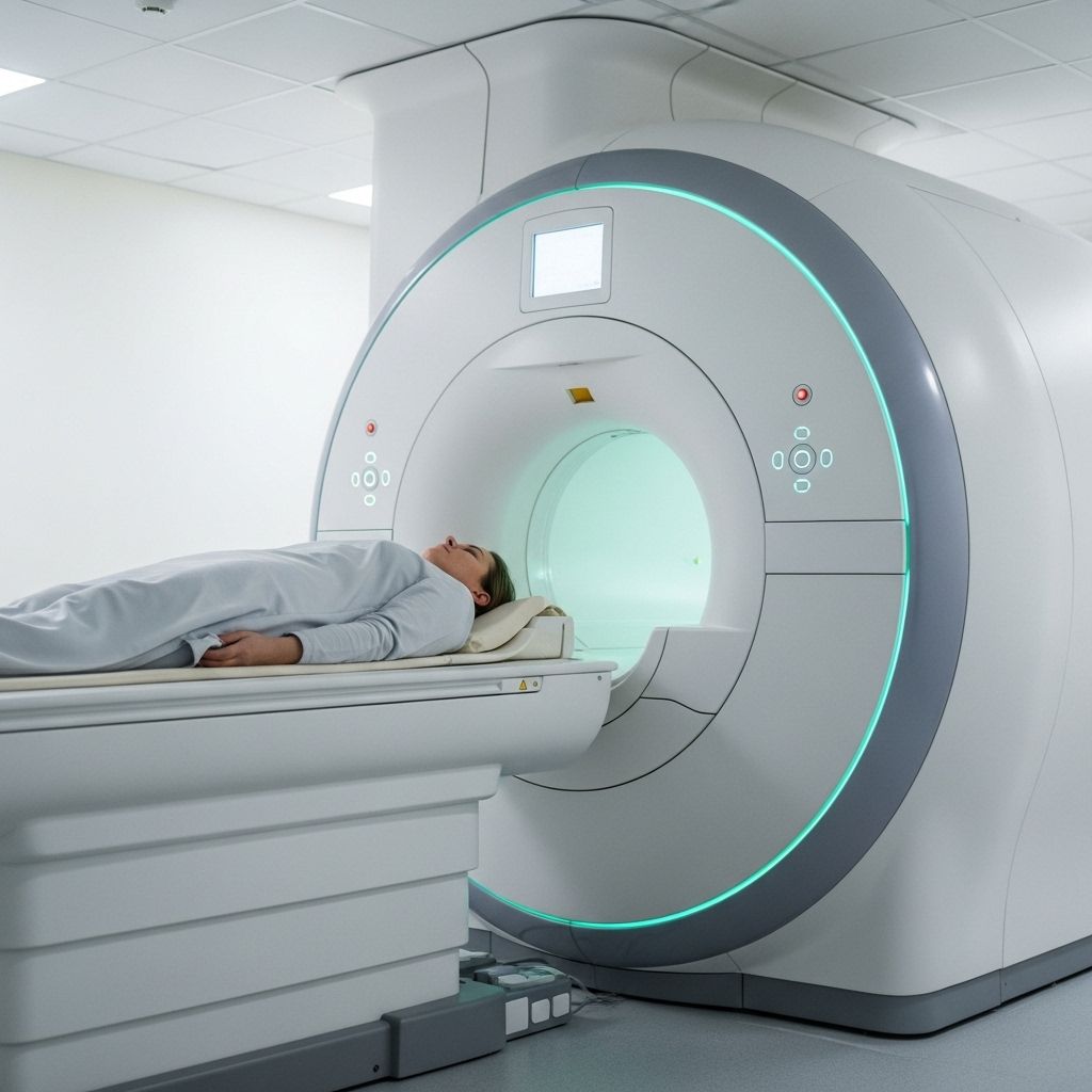
Magnetic Resonance Imaging (MRI): What to Expect, Uses, and Safety
Learn how MRI scans work, when they are used, what to expect, and essential safety facts before your imaging appointment.

Magnetic Resonance Imaging, commonly known as MRI, is a sophisticated, non-invasive imaging technique used by medical professionals to view detailed images of internal structures within the body. MRI technology plays a crucial role in diagnosing and monitoring a variety of conditions, leveraging powerful magnets, radio waves, and advanced computer processing to create cross-sectional images without using ionizing radiation like X-rays or CT scans.
What Is Magnetic Resonance Imaging (MRI)?
MRI is an imaging modality that utilizes a large magnet, radiofrequency energy, and computer technology to generate high-resolution images of organs, tissues, and even bones. Unlike traditional X-rays or computed tomography (CT), MRI does not involve exposure to potentially harmful radiation.
- Non-invasive: MRI scans do not require surgery or insertion of devices into the body.
- Detailed images: It provides clear, high-contrast images, especially of soft tissues such as the brain, muscles, joints, and internal organs.
- No ionizing radiation: It uses magnetic fields and radio waves instead of X-rays.
How Does an MRI Work?
The technology behind MRI revolves around magnetic fields and radio waves interacting with the body’s water molecules. Here’s a brief overview:
- Powerful magnetic field: A patient lies within a large cylindrical magnet, which aligns the hydrogen protons in the body.
- Radiofrequency pulses: The scanner transmits radio waves, temporarily knocking the protons out of alignment.
- Signal capture and processing: When the radiofrequency is turned off, protons realign, releasing signals that are captured by the scanner and converted into images by a computer.
This process allows for accurate visualization of internal anatomy and detection of abnormalities.
Reasons for MRI Scanning
Physicians order MRI scans to diagnose, plan treatment, or monitor many different health conditions. MRI has become a cornerstone in various medical specialties due to its clarity and versatility.
Common Medical Indications for an MRI
- Neurological conditions: MRI is essential for evaluating the brain and spinal cord for tumors, multiple sclerosis, stroke, aneurysms, or inflammation.
- Bone and joint issues: Used to diagnose torn ligaments, cartilage injury, bone infections, and tumors.
- Cardiovascular assessments: MRI can detect heart structure abnormalities, heart disease, or blood vessel problems.
- Abdominal and pelvic imaging: Provides detailed images of the liver, kidneys, spleen, uterus, and prostate, helping diagnose tumors, infections, and other organ-specific diseases.
- Cancer diagnosis and staging: MRI is often employed to pinpoint tumor location, measure size, and plan interventions.
Different Types of MRI Exams
The versatility of MRI means it is adapted to image different parts of the body and answer specific clinical questions. Some specialized MRI approaches include:
- Brain and spinal cord MRI: For neurological evaluation.
- MRI angiography (MRA): Focused on imaging blood vessels.
- Musculoskeletal MRI: For joints, cartilage, and soft tissue analysis.
- Cardiac MRI: To visualize heart structures and function.
- Functional MRI (fMRI): Used in research to observe brain activity in real time.
Some MRI scans require a special dye, or contrast agent (usually gadolinium), injected through a vein to highlight specific tissues or blood vessels.
Preparing for an MRI Procedure
Preparation is essential to ensure both the safety and effectiveness of the MRI scan. Patients should always follow their care team’s specific instructions, but general guidelines typically include:
- Review all prior instructions: Some exams require fasting or other special preparation.
- Complete screening forms: Patients are asked to fill out a screening form to identify any risk factors, such as metal implants, pacemakers, or previous surgeries.
- Remove metal objects: All jewelry, glasses, dentures, and anything containing metal must be removed before entering the scan room.
Who Should Inform Their Care Team?
- Pregnant or possibly pregnant patients
- Those who have surgically implanted medical devices (e.g., pacemakers, artificial joints, cochlear implants)
- Individuals with metal fragments or bullets in the body
- People with allergies to contrast agents
- Anyone with a history of kidney disease (since certain contrast agents are processed by the kidneys)
What Happens During the MRI Procedure?
The MRI experience can vary depending on the part of the body being scanned and whether contrast dye is used, but most procedures follow similar steps:
- Arrival and check-in: The care team will review the screening questionnaire and answer questions.
- Changing: Patients may be asked to change into a hospital gown to prevent any metal interference.
- Positioning: The technologist helps the patient lie on the MRI table, ensuring comfort and correct alignment.
- Entering the scanner: The table slides the patient into the cylindrical scanning chamber. Patients must remain very still for best image quality.
- Imaging: The MRI machine generates loud tapping or knocking sounds during scanning; earplugs or headphones are usually provided to reduce discomfort.
- Contrast administration (if needed): For certain scans, a contrast agent may be given through an IV during the exam to enhance image detail.
- Communication: Patients are in constant touch with the technologist through a microphone. Most scanners allow patients to signal if they require assistance.
Depending on the region being studied, MRI exams may take approximately 30 to 90 minutes. The body part being scanned must stay completely still during imaging sequences to avoid blurring. Breath-holding instructions may be given for abdominal or chest scans.
What to Expect After the MRI
After the scan:
- Patients are generally free to resume normal activities immediately.
- If a contrast agent was used, instructions about drinking fluids may be provided to help flush the dye out of the body.
- There are typically no restrictions on eating or physical activity post-procedure.
- The MRI technologist will not discuss scan results with the patient; instead, a radiologist (specialist in reading imaging studies) will review the images and send a detailed report to the referring provider.
MRI Safety: What You Should Know
While MRI is generally considered extremely safe, the powerful magnetic field means that some individuals and items are strictly prohibited around the scanner.
Important MRI Safety Guidelines
- Metal and electronic implants: Notify your care team if you have pacemakers, aneurysm clips, cochlear implants, infusion pumps, nerve stimulators, or any embedded electronic device.
- Screening protocols: Every patient undergoes rigorous screening to prevent unsafe exposure to magnetic forces.
- Four-zone concept: Many MRI facilities use a safety system, designating four zones that control access from the public area to the secure scanning room.
- Contrast risks: Most people tolerate gadolinium contrast well, but those with severe kidney disease should avoid it due to rare complications.
- Pregnancy: Current research suggests that MRI (without contrast) is safe in pregnancy, but always discuss any questions with your doctor.
Special Technology and Innovations
- Autonomous MRI (AMRI): Artificial intelligence and autonomous workflow advances are making MRI more accessible and efficient, with cloud-based protocols and user-guided voice assistants simplifying exams for patients and clinicians alike.
- Research advances: New algorithms are being developed, like DeepSTI, to improve brain imaging using fewer scans, reducing the burden for patients and increasing diagnostic clarity for neurological diseases.
- Magnetic susceptibility mapping: Advanced techniques can now provide detailed maps of brain tissue characteristics, enhancing ability to diagnose and monitor conditions such as multiple sclerosis and Alzheimer’s disease.
Who Should Not Have an MRI?
The following situations may prevent or require extra caution for an MRI:
- Having a non-MRI-compatible pacemaker or implanted defibrillator
- Certain metallic implants, ferromagnetic aneurysm clips, or cochlear implants
- Metal fragments in the eye or body, especially if not removable
- Severe kidney disease (if contrast is required)
- Allergic history to MRI contrast agents
- Early pregnancy without clear medical necessity
Always disclose your complete medical and surgical history. In some cases, alternative imaging tests (such as ultrasound or CT) may be recommended if MRI is contraindicated.
Frequently Asked Questions (FAQs)
How long does an MRI scan take?
Most MRI procedures last between 30–90 minutes, depending on the area being studied and whether contrast dye is used.
Can I move during the MRI?
Remaining absolutely still is necessary during each scan sequence. Any movement can blur or distort the images, requiring repeated scans.
Is MRI safe for children?
Yes, MRI is routinely performed for children. Sedation or anesthesia may be used for young or anxious patients to help them stay still during the procedure.
Are there side effects from MRI contrast?
Gadolinium-based contrast agents are usually very safe. Mild allergic reactions are rare. People with severe kidney disease should avoid them to prevent a rare complication called nephrogenic systemic fibrosis.
Is there any risk from the magnetic field?
The magnetic field does not pose a health risk, but it can affect heart devices, metal implants, or items in or on the body. Strict safety screenings prevent adverse events.
What if I am claustrophobic or anxious?
Tell your healthcare team before your MRI appointment. Options may include open MRI scanners, calming music, or medication to reduce anxiety. In some cases, sedation can be arranged if needed.
Tips for a Successful MRI Scan
- Arrive early to complete forms and allow time for questions.
- Wear comfortable, loose-fitting clothes without metallic fasteners. You may also be provided a gown.
- Leave valuables and electronic devices at home.
- Follow all instructions from your MRI technologist regarding eating, drinking, and medication use prior to the appointment.
- If you have a history of claustrophobia, inform staff in advance so accommodations can be made.
Summary Table: MRI Key Facts
| Aspect | Details |
|---|---|
| Type of Test | Non-invasive, cross-sectional imaging using magnetic fields |
| Primary Uses | Diagnosis of neurological, musculoskeletal, cardiovascular, and abdominal conditions |
| Preparation Required | Screening for metal, fasting for some exams, removal of metallic items |
| Duration | 30–90 minutes (varies by scan type) |
| Contrast Use | Required for some studies (e.g., tumors, blood vessels) |
| Radiation | None (uses magnetic fields and radio waves) |
| Risks | Metal and device complications, rare allergic reactions to contrast |
Conclusion
Magnetic Resonance Imaging (MRI) stands out as a vital, versatile technology in the diagnostic process for countless medical conditions. By providing high-definition images of internal organs and tissues without radiation, MRI empowers healthcare professionals to offer accurate diagnoses and monitor complex diseases safely. Understanding what to expect before, during, and after your MRI scan helps reduce anxiety and ensures the best possible experience and outcome.
References
- https://hub.jhu.edu/2023/12/14/brain-imagining-less-data/
- https://pure.johnshopkins.edu/en/publications/autonomous-magnetic-resonance-imaging
- http://jhrad.com/mri/policy_SAFETY_2020.pdf
- https://www.youtube.com/watch?v=z89KOf3WIrk
- https://pure.johnshopkins.edu/en/publications/accessible-magnetic-resonance-imaging-a-review
- https://jpmsonline.com/article/jpms-volume-1-issue-1-pages29-31-er/
Read full bio of Sneha Tete


