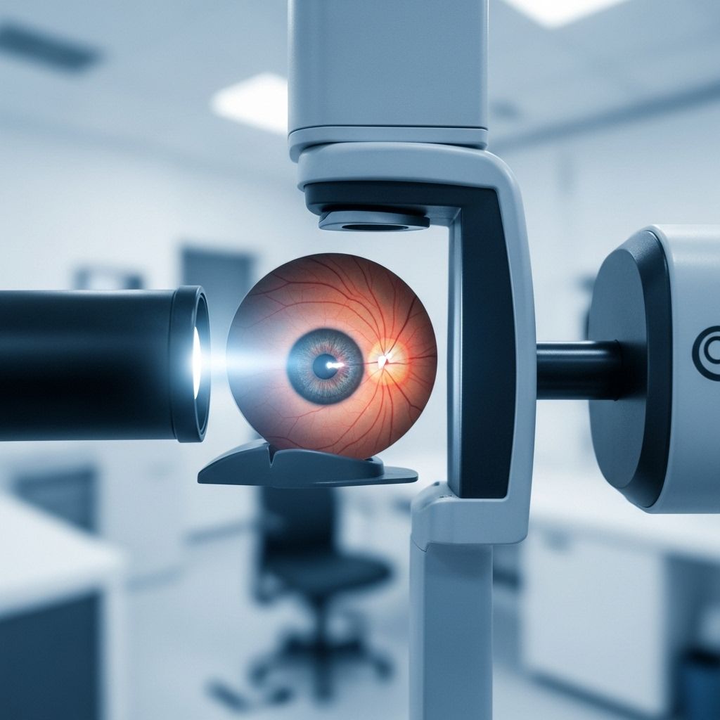
Macular Degeneration Testing: Methods, Diagnosis, and What to Expect
Learn about the comprehensive tests and procedures used to diagnose macular degeneration and how you can monitor your eye health.

Macular degeneration is a progressive eye disease affecting the macula, the central part of the retina responsible for sharp, straight-ahead vision. Early detection is crucial for preserving vision and tailoring effective treatment strategies. This article covers the essential exams, advanced imaging techniques, genetic tests, and self-monitoring methods used for diagnosing and tracking macular degeneration.
What is Macular Degeneration?
Macular degeneration refers to conditions that damage the central retina, resulting in blurry or distorted central vision. It commonly relates to age-related changes (AMD) but can also be caused by inherited retinal diseases or other medical conditions. Symptoms often begin with subtle changes, such as difficulty reading or noticing wavy lines, and can progress to significant vision impairment in advanced stages.
How Is Macular Degeneration Diagnosed?
Diagnosis typically starts with a comprehensive eye exam performed by an ophthalmologist or optometrist. This exam assesses both vision and eye health, incorporating several key tests and procedures:
- Medical and Family History Review: Understanding risk factors and family predisposition.
- Pupil Dilation: Eye drops expand the pupil for a clearer view of the retina.
- Back of Eye Examination: Specialists look for the presence of drusen (yellow retinal deposits), retinal atrophy, and other signs of macular disease.
- Vision Field Testing: The Amsler grid is used to check for central vision distortion.
Common Symptoms Leading to Diagnosis
- Blurriness or distortion in central vision
- Difficulty with detail-oriented tasks
- Wavy or faded lines noticed during self-testing
- Central blind spots (especially as the disease progresses)
Key Tests and Imaging for Macular Degeneration
Beyond the basic eye exam, several advanced imaging techniques allow eye care professionals to visualize the retina’s structure and blood vessels in detail:
Optical Coherence Tomography (OCT)
- Uses light waves to capture cross-sectional retina images.
- Identifies thinning, swelling, or fluid buildup indicative of macular change.
- Painless and noninvasive; completed in 5–10 minutes.
Visible Light OCT
- An emerging technology using visible rather than near-infrared light.
- May offer improved image contrast for certain retinal conditions.
OCT Angiography (OCTA)
- Provides blood vessel visualization without injectable dye.
- Useful for assessing vascular changes in wet macular degeneration.
Fluorescein Angiography
- A fluorescent dye is injected into a vein; photographs track dye through retinal vessels.
- Highlights leaking blood vessels typical of advanced or wet macular degeneration.
Indocyanine Green Angiography
- Uses injected green dye to enhance visualization of deeper blood vessels.
- Often combined with fluorescein angiography for complex cases.
Fundus Autofluorescence Imaging
- Noninvasive technique capturing the retina’s natural cell fluorescence.
- Assists in diagnosing and monitoring various macular conditions.
Genetic Testing
Hereditary forms of macular degeneration may require genetic testing. Your doctor may recommend a simple blood test if symptoms suggest an inherited retinal condition, such as Best disease or Stargardt’s disease. Genetic analysis can confirm diagnosis and inform family members about potential risks.
Self-Monitoring: The Amsler Grid
The Amsler grid is a widely recommended at-home screening tool developed by Marc Amsler in 1945. It helps individuals detect and monitor changes to their central vision between regular doctor visits.
Instructions for Using the Amsler Grid:
- Test in a well-lit room using prescribed glasses if needed.
- Hold the grid about 12–14 inches from your eyes.
- Cover one eye; focus with the open eye on the central dot.
- Answer the self-assessment questions provided with the grid (record any distortions, faded spots, or wavy lines).
- Repeat the test with the other eye.
If any abnormalities are noticed, report them to your eye doctor. The Amsler grid is not a substitute for regular professional evaluation—it is best used for ongoing self-monitoring.
Interpreting Test Results and Next Steps
- Mild cases of dry age-related macular degeneration (AMD): Often monitored with annual or biannual checkups. Self-testing with the Amsler grid is advisable.
- Active or advanced wet AMD: May require monthly injections to control abnormal blood vessel growth and leakage. Treatment frequency can adjust based on progress.
Regardless of the diagnosis, following your eye doctor’s recommendations for monitoring and treatment is crucial for preserving sight.
What Can Be Mistaken for Macular Degeneration?
Macular degeneration shares symptoms with several other eye conditions. Differential diagnosis is necessary to rule out:
- Retinal detachment
- Epiretinal membrane syndrome
- Diabetic retinopathy
- Central serous retinopathy
Comprehensive Table: Macular Degeneration Tests and Their Purpose
| Test | Method | Purpose | Invasiveness |
|---|---|---|---|
| Comprehensive Eye Exam | Visual acuity & dilated retinal assessment | Initial detection, overall eye health | Noninvasive |
| Optical Coherence Tomography (OCT) | Cross-sectional retina imaging | Detects swelling, thinning, fluid, structure changes | Noninvasive |
| OCT Angiography (OCTA) | Retinal blood vessel imaging | Detects vascular abnormalities | Noninvasive |
| Fluorescein Angiography | Injectable dye photography | Highlights leaking blood vessels, wet AMD | Minimally invasive |
| Indocyanine Green Angiography | Green dye vessel imaging | Examines deeper blood vessels; complex cases | Minimally invasive |
| Fundus Autofluorescence | Cell fluorescence imaging | Assesses retinal health and macular changes | Noninvasive |
| Genetic Testing | Blood sample analysis | Detects inherited retinal diseases | Minimally invasive |
| Amsler Grid | In-home self-assessment | Monitors central vision distortion | Noninvasive |
Frequently Asked Questions About Macular Degeneration
Can an optometrist detect early signs of macular degeneration?
Yes, optometrists can identify early signs—especially during a comprehensive eye exam. They may refer you to a retina specialist for confirmation and ongoing treatment.
What is the first indication of macular degeneration?
Most people first notice blurriness or distortion in their central vision. Any changes to vision should be promptly discussed with an eye care professional.
What can be mistaken for macular degeneration?
Other medical eye conditions, such as retinal detachment or epiretinal membrane syndrome, may mimic the symptoms of macular degeneration. Only detailed examination can confirm or rule out these diagnoses.
Do I need a comprehensive exam if one Amsler grid response is abnormal?
Yes, any abnormality on the Amsler grid warrants a professional examination. While useful as a monitoring tool, the grid cannot diagnose underlying diseases, and abnormal results require follow-up with an eye doctor.
Are macular degeneration tests painful?
The majority of diagnostic tests, including the comprehensive eye exam and most imaging, are painless. Angiography involves an injectable dye but is generally well tolerated.
How often should I have my eyes checked for macular degeneration?
For patients with risk factors or diagnosed dry AMD, annual or twice-yearly checkups are recommended. Individuals with wet AMD may require monthly follow-ups, especially when receiving treatments.
Can genetic testing help my family?
Genetic testing may clarify whether a hereditary form of macular degeneration is present, helping family members understand their own risk.
Key Takeaways
- Early detection of macular degeneration relies on comprehensive eye exams combined with advanced imaging and occasional genetic testing.
- The Amsler grid is a practical self-monitoring tool but does not replace professional evaluation.
- A variety of tests, both noninvasive and minimally invasive, help differentiate types and severity of macular degeneration.
- Timely intervention and adherence to medical advice can slow progression and preserve vision.
References
- https://www.healthline.com/health/how-is-macular-degeneration-diagnosed
- https://www.wandmeyes.com/eye-care-services/eye-disease-management/treating-macular-degeneration/take-the-macular-degeneration-test/
- https://www.mayoclinic.org/diseases-conditions/dry-macular-degeneration/diagnosis-treatment/drc-20350381
- https://www.brightfocus.org/resource/amsler-grid-eye-test/
- https://www.medicalnewstoday.com/articles/152105
- https://medlineplus.gov/maculardegeneration.html
- https://stanfordhealthcare.org/medical-conditions/eyes-and-vision/macular-degeneration/diagnosis.html
Read full bio of Sneha Tete


