Inferior Vena Cava (IVC) Filter Placement: Procedure, Benefits, and Considerations
Learn about the use, process, safety, and removal of IVC filters for preventing life-threatening pulmonary embolism.

Inferior Vena Cava (IVC) Filter Placement
Inferior vena cava (IVC) filter placement is a vital procedure used to prevent life-threatening complications that arise from blood clots traveling to the lungs. This article provides an extensive review of IVC filter placement, highlighting its purpose, when it’s recommended, how the procedure is performed, potential risks, and key considerations for patients.
What Is an Inferior Vena Cava (IVC) Filter?
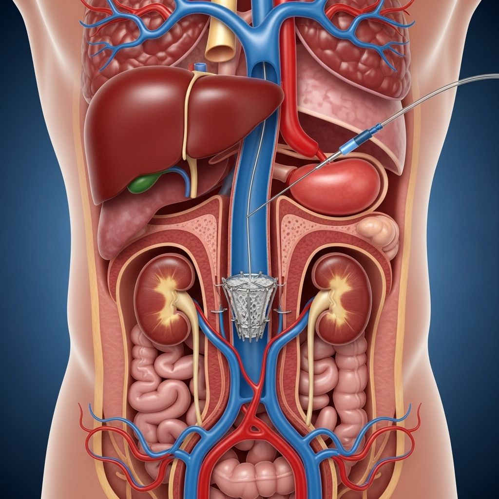
The inferior vena cava is the largest vein in the body, running through the abdomen and transporting blood from the lower body back to the heart. An IVC filter is a small, wire, umbrella-like device placed in this vein to catch blood clots before they can move to the lungs and cause a pulmonary embolism (PE), which can be fatal if untreated .
Why Might You Need an IVC Filter?
The main reason for IVC filter placement is to reduce the risk of pulmonary embolism in people who:
- Have or are at risk for deep vein thrombosis (DVT)—a blood clot in the deep veins, usually in the legs.
- Cannot take anticoagulation (blood-thinning) medications because of side effects, allergies, or other medical conditions.
- Continue to develop clots despite taking blood thinners.
- Are undergoing procedures that temporarily increase the risk of clots, such as major surgeries or trauma.
- Have experienced bleeding complications from anticoagulant therapy or have a high risk of such complications.
In most situations, anticoagulants remain the first line of defense against DVT and PE, but IVC filters are crucial when these medications are not an option or are not effective .
How Does an IVC Filter Work?
An IVC filter traps large clots that break loose from veins in the legs or pelvis, preventing them from traveling upward to the lungs. The device is positioned so it allows normal blood flow, but its struts catch and hold clots until they dissolve naturally or are treated .
Types of IVC Filters
There are two primary types of IVC filters:
- Permanent filters: Designed to remain in the IVC indefinitely. Used when the risk for clots is ongoing or permanent.
- Retrievable (temporary) filters: Intended for short-term protection and can be removed when the risk subsides. Most newer filters are designed with this option.
Preparing for IVC Filter Placement
The procedure is usually scheduled in advance, except in emergencies. Preparation steps often include:
- Providing a detailed medical history, including all medications, allergies (particularly to contrast dye, anesthetics, or latex), and prior procedures.
- Discussing current blood thinners or other medications with the healthcare team.
- Fasting for several hours before the procedure if instructed.
- Arranging for transportation home, as sedation may be used.
How Is an IVC Filter Placed?
The placement of an IVC filter is generally a minimally invasive, image-guided procedure performed by an interventional radiologist or vascular surgeon. Here is a step-by-step overview:
- Patient Positioning and Site Preparation
A patient lies flat on their back. The neck or groin (the site of vein access) is cleaned thoroughly and covered with a sterile drape. - Local Anesthesia
A local anesthetic is injected at the puncture site to numb the area. Moderate sedation may also be provided to keep the patient comfortable. - Venous Access
A needle is used to access a large vein, typically in the neck or groin. - Catheter Insertion
A catheter (thin flexible tube) is inserted through the vein and advanced to the IVC using real-time X-ray (fluoroscopy) guidance. - Contrast Dye Injection
Contrast dye is injected through the catheter to visualize the IVC and confirm placement accuracy. - Filter Deployment
The filter is passed through the catheter and deployed in the desired location within the vena cava. - Catheter Removal and Bandaging
The catheter is removed, and the puncture site is compressed to stop any bleeding. A bandage is applied over the area.
The procedure typically takes about 30–60 minutes. Most patients remain awake but relaxed, experiencing only minor discomfort at the needle insertion site .
After the Procedure: What to Expect
Following IVC filter placement, patients are monitored for several hours:
- The insertion site is checked for bleeding or infection.
- Vital signs (heart rate, blood pressure, etc.) are monitored.
- Pain and discomfort at the site are usually minimal and short-lived.
- Most people are discharged the same day with recovery instructions and may resume normal activities within a day or two.
It is crucial to attend all follow-up appointments, regardless of whether the filter is intended to be permanent or temporary. These appointments help determine if filter removal is appropriate and check for any signs of complications .
Risks and Complications
While IVC filter placement is generally safe, potential risks and complications may include:
- Bleeding or hematoma at the puncture site.
- Infection at the puncture site.
- Reaction to contrast dye (rare).
- Filter migration or misplacement.
- Filter fracture or breakage.
- Blood vessel injury or perforation.
- Development of a new blood clot at the filter site.
- Long-term risks if the filter remains in place unnecessarily, such as IVC narrowing or occlusion.
Discuss any concerns about risks with a healthcare provider prior to the procedure .
IVC Filter Removal
Routine Filter Removal
Most filters are designed for retrieval once the risk of thrombosis or embolism has lessened. Reasons to remove the filter include:
- The original risk (such as surgery or trauma) has resolved.
- The patient can safely resume anticoagulation therapy.
- Potential device-related complications arise.
Removal is a minimally invasive outpatient procedure, similar to placement. A catheter is guided to the filter, which is then captured—usually with a device called a snare—collapsed, and withdrawn. Additional imaging checks confirm full removal. Routine removal usually takes 30–60 minutes and uses local anesthesia and moderate sedation .
Complex Filter Removal
In some cases, an IVC filter may become embedded in the vein wall or fracture into pieces. These situations require specialized techniques, possibly including CT scans for planning, larger or dual catheters, snare devices, forceps, or laser-assisted removal. Such procedures can be longer (up to two hours or more) and may require general anesthesia. Referral to a highly experienced center is often necessary for complex cases .
Recovery and Aftercare
Key points for a smooth recovery after IVC filter placement or removal:
- Follow all discharge instructions provided by your care team.
- Monitor the access site for signs of bleeding, swelling, redness, or infection.
- Avoid heavy lifting or strenuous activity for 24–48 hours.
- Report any new pain, numbness, shortness of breath, or swelling to your provider immediately.
- Adhere to prescribed medications, including blood thinners if recommended.
- Attend all scheduled follow-up appointments for imaging and evaluation.
Frequently Asked Questions (FAQs)
Q: How long does IVC filter placement take?
A: The procedure typically lasts about 30 to 60 minutes, but complex cases may take longer.
Q: Is IVC filter placement painful?
A: Minor discomfort or a sting from local anesthesia is possible, but most people feel little pain during the procedure due to sedation and numbing medications.
Q: Can I return to normal activities quickly?
A: Most patients are able to resume normal activities within a day or two, although strenuous activity should be avoided for the first 24–48 hours.
Q: Are IVC filters permanent implants?
A: Some filters are permanent, but many are designed to be temporary and removable once the clotting risk has subsided and it’s safe to discontinue.
Q: What are the signs of a complication?
A: Watch for persistent pain, swelling, redness or bleeding at the insertion site, fever, sudden shortness of breath, chest pain, or leg swelling. Seek immediate medical attention if any of these occur.
SEO Table: Quick Facts on IVC Filter Placement
| Aspect | Details |
|---|---|
| Purpose | Prevent pulmonary embolism by trapping blood clots |
| Indications | Patients with DVT/PE and unable to use anticoagulants or with recurrent clots |
| Placement Site | Inferior vena cava in the abdomen |
| Procedure Length | 30–60 minutes (can be longer if complex removal needed) |
| Anesthesia | Local anesthesia, moderate sedation, occasionally general anesthesia |
| Risks | Bleeding, infection, filter migration, vessel damage, new clot |
| Recovery | Same day discharge; resumption of activity within 24–48 hours |
| Filter Removal | Recommended when clot risk subsides; uses similar technique as placement |
Key Reminders
- IVC filter placement is not a substitute for anticoagulation except in specific cases.
- Timely removal of temporary filters is important to reduce risk of long-term complications.
- Always maintain close follow-up with your healthcare provider after the procedure.
Understanding the use and indications for IVC filter placement enables patients and families to make informed decisions and participate actively in their own care. If you or a loved one is considering or has been recommended for an IVC filter, discuss any questions or concerns with your medical team to ensure the safest, most effective treatment plan.
References
- https://rad.uw.edu/sections/interventional-radiology/procedures/inferior-vena-cava-ivc-filters
- https://vein.stonybrookmedicine.edu/treatments/inferior-vena-cava-filters
- https://www.radiologyinfo.org/en/info/venacavafilter
- https://www.azuravascularcare.com/medical-services/ivc-filter-placement-procedure-removal/
- https://my.clevelandclinic.org/health/treatments/17609-vena-cava-filters
- https://pmc.ncbi.nlm.nih.gov/articles/PMC8175103/
- https://www.yalemedicine.org/conditions/ivc-filter-placement-and-removal
- https://www.mskcc.org/cancer-care/patient-education/ivc-filter-placement
Read full bio of Sneha Tete


