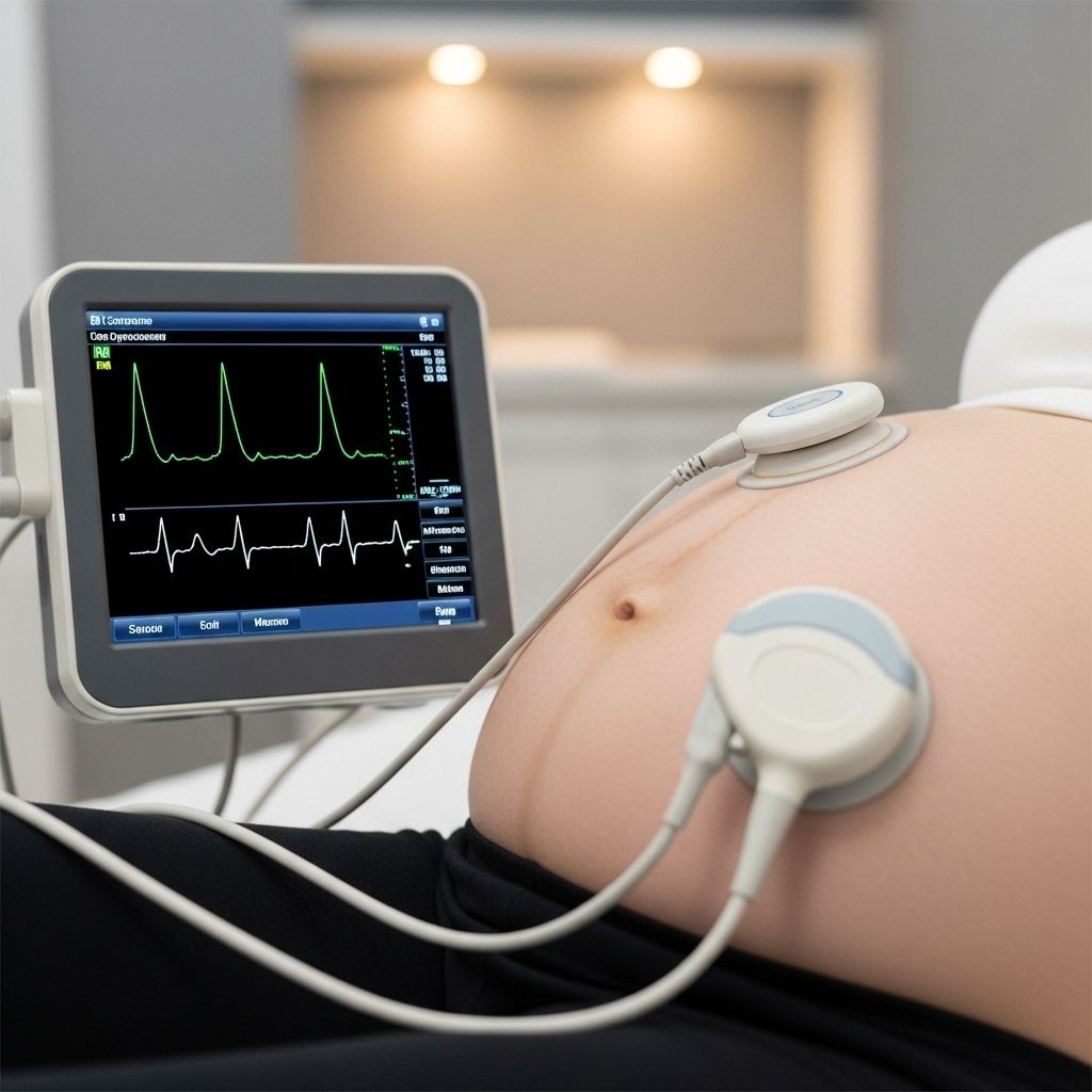
Fetal Heart Monitoring: Methods, Purpose, and Interpretation
Understanding fetal heart monitoring: procedures, techniques, and their vital role in safeguarding prenatal health.

Fetal heart monitoring is a critical diagnostic tool used during pregnancy and labor, providing vital information about the well-being of the unborn baby. By tracking the fetal heart rate and rhythm, healthcare providers can assess the fetus’s health and respond promptly to any distress or abnormality. This article explores fetal heart monitoring techniques, procedures, interpretation, and addresses frequently asked questions to help expectant parents understand its importance.
What is Fetal Heart Monitoring?
Fetal heart monitoring refers to the measurement and analysis of the baby’s heart rate inside the womb. Its primary goal is to evaluate the fetus’s well-being, identify possible distress, and help guide clinical decisions during pregnancy and labor.
- Checks fetal heart rate and rhythm to identify increases, decreases, or abnormal patterns.
- Provides real-time data on the fetus’s response to intrauterine conditions and uterine contractions.
- Enables providers to intervene quickly in cases of fetal compromise, such as reduced oxygen supply.
Why is Fetal Heart Monitoring Performed?
Fetal heart monitoring serves several purposes:
- Ensures fetal well-being during pregnancy and labor.
- Detects signs of fetal distress or lack of oxygen.
- Aids decision-making for interventions, like a cesarean delivery, if emergencies arise.
- Assesses how the fetus tolerates labor and contractions.
The average normal fetal heart rate ranges from 110 to 160 beats per minute. Significant changes can signal conditions requiring immediate medical attention.
How is Fetal Heart Monitoring Performed?
There are two main methods for monitoring the fetal heart rate: external (non-invasive) and internal (invasive).
External Fetal Heart Monitoring
- Uses a fetoscope (special stethoscope) or Doppler device.
- Devices detect and transmit the fetal heartbeat sounds or signals to a monitor.
- Can be intermittent or continuous, depending on maternal and fetal health.
- During labor, an external ultrasound transducer is placed on the mother’s abdomen using an elastic belt to keep it secure.
- A second device, called an external tocodynamometer, may be placed over the abdomen to record contractions.
- The fetal heartbeat is displayed on a screen and recorded on a printed strip for analysis.
External Monitoring Procedure
- A clear gel is applied to the abdomen to conduct sound waves effectively.
- The transducer is placed and moved until the fetal heart rate is located.
- Continuous monitoring involves securing the transducer with a belt and recording heart rate data.
- After the procedure, straps and gel are removed.
Internal Fetal Heart Monitoring
- Performed if more accurate readings are required, such as for high-risk pregnancies or when external monitoring is challenging.
- Requires that the amniotic sac (bag of waters) be ruptured and the cervix partially dilated.
- A fetal scalp electrode is gently attached to the baby’s scalp through the mother’s cervix.
- The electrode transmits heart rate signals to a monitor via a wire.
- After delivery, the electrode is removed and the baby’s scalp is examined for injury.
Internal Monitoring Procedure
- The expectant mother undresses and lies on the labor bed as for a pelvic exam.
- A gloved provider checks for cervical dilation.
- If membranes are intact, the provider may rupture them using an instrument.
- The electrode guide is gently inserted into the vagina and the electrode attached to the fetal skin.
- The electrode is connected to monitoring equipment and secured with a band around the thigh.
- Electrode is removed once the baby is born.
Types of Fetal Heart Monitoring
| Method | How It Works | When Used | Advantages | Limitations |
|---|---|---|---|---|
| External Monitoring | Transducer placed on abdomen, uses Doppler ultrasound | Routine checks, low-risk pregnancies | Non-invasive, no risk for baby | May lose signal if mother moves, less accurate |
| Internal Monitoring | Electrode attached to fetal scalp, signals sent to monitor | High-risk pregnancies, difficult external monitoring | Accurate, reliable signals | Requires ruptured membranes, some risk (infection, injury) |
Intermittent vs. Continuous Monitoring
- Intermittent: Heart rate measured at regular intervals (e.g., every 30 minutes for low-risk, every 15 minutes for high-risk patients).
- Continuous: Heart rate is observed without interruption throughout labor, typically reserved for high-risk cases or when abnormalities are detected during intermittent checks.
- Intermittent monitoring can be effective and is recommended by organizations such as ACOG (American College of Obstetricians and Gynecologists) for most low-risk pregnancies.
Preparation for Fetal Heart Monitoring
- No special dietary restrictions; most patients continue normal activities unless advised differently by their provider.
- Prior to hospital-based monitoring, patients typically change into a gown and lie in a comfortable position.
- For internal monitoring, the provider will ensure that conditions (ruptured membranes, cervical dilation) are appropriate before proceeding.
Risks of Fetal Heart Monitoring
- External monitoring: No physical risks as the procedure is non-invasive.
- Internal monitoring:
- Small risk of infection at the electrode site on the baby’s scalp.
- Possible bruising or minor lacerations at the attachment site.
- Requires ruptured membranes and partial cervical dilation, which may not be suitable for all patients.
After Fetal Heart Monitoring
- External: Resume normal activities immediately.
- Internal: Provider examines the baby’s scalp for possible infection, bruising, or laceration and cleans the site if needed.
- Providers may give additional care instructions depending on individual situations.
How Are Fetal Heart Rate Patterns Interpreted?
Interpretation of fetal heart monitoring requires careful evaluation of baseline rate, rhythm, variability, and patterns:
- Baseline FHR (Fetal Heart Rate): The average heartbeat per minute, typically 110–160 bpm.
- Variability: Fluctuations in heart rate; good variability suggests healthy fetal oxygenation.
- Accelerations: Temporary increase in heart rate; usually reassures well-being.
- Decelerations: Drops below baseline; may signal distress if prolonged or deep.
| Pattern | What It May Indicate |
|---|---|
| Normal | Well-oxygenated, healthy fetus |
| Bradycardia (slow) | Possible fetal distress, cord compression, medications |
| Tachycardia (fast) | Might indicate maternal fever, infection, or prematurity |
| Absent/minimal variability | May suggest fetal hypoxia or drugs affecting baby |
| Late decelerations | Pertinent for fetal compromise, possible need for intervention |
| Variable decelerations | Often indicates cord compression, usually transient but may be concerning |
When Is Fetal Heart Monitoring Typically Used?
- Throughout pregnancy during prenatal appointments to check fetal health.
- During labor for both low-risk and high-risk pregnancies.
- Required for certain situations, including:
- Mothers with medical conditions (e.g., hypertension, diabetes)
- Suspected fetal growth restriction
- Preterm labor
- Induced labor or prolonged labor
- Use of epidural analgesia
- Multiple gestation pregnancies
Frequently Asked Questions About Fetal Heart Monitoring
Q: Is fetal heart monitoring painful?
A: External monitoring is painless, though the transducer and belt may feel tight. Internal monitoring may cause minor discomfort during electrode placement but should not be painful overall.
Q: Are there any risks to fetal heart monitoring?
A: External monitoring carries no known risks. Internal monitoring carries a slight risk of infection, bruising, or minor injury to the baby’s scalp, which is generally rare and managed quickly by medical staff.
Q: How long does fetal heart monitoring take?
A: Monitoring duration varies: prenatal appointments may involve brief checks, while labor could require continuous monitoring depending on maternal/fetal risk factors.
Q: Can I move around during fetal heart monitoring?
A: During intermittent external monitoring, movement is generally allowed, but during continuous monitoring (especially with internal electrodes), you may be asked to remain in bed or limit movement.
Q: What happens if an abnormal heart rate is detected?
A: Providers will assess for possible causes (such as cord compression, low oxygen), take corrective actions (repositioning, oxygen for mother, stopping offending medications), and may recommend emergency intervention if fetal distress continues.
Key Points and Summary
- Fetal heart monitoring is a vital safety measure to assess fetal health during pregnancy and labor.
- External methods are non-invasive and suitable for most routine monitoring; internal methods are reserved for high-risk or when accurate readings are required.
- Patterns in heart rate—especially baseline rate, variability, accelerations, and decelerations—guide provider decisions and interventions.
- Both monitoring methods are regarded as safe with minimal risk, and preparation/aftercare are straightforward and non-burdensome for most patients.
References
- https://www.stanfordchildrens.org/en/topic/default?id=fetal-monitoring-90-P02448
- https://www.urmc.rochester.edu/encyclopedia/content?contentid=P07776&contenttypeid=92
- https://www.aafp.org/pubs/afp/issues/1999/0501/p2487.html
- https://my.clevelandclinic.org/health/diagnostics/23464-fetal-heart-rate-monitoring
- https://www.acog.org/womens-health/faqs/fetal-heart-rate-monitoring-during-labor
- https://www.ncbi.nlm.nih.gov/books/NBK589699/
- https://www.youtube.com/watch?v=aSoWQbE4MIQ
- https://www.webmd.com/baby/pregnancy-fetal-heart-monitoring
Read full bio of medha deb


