Electroencephalogram (EEG): Purpose, Procedure, Types, and What to Expect
Discover how EEG tests monitor brain activity, why they're ordered, what to expect during the procedure, and how results guide neurological care.

An electroencephalogram (EEG) is a widely used and non-invasive test that detects electrical activity in your brain using small sensors placed on the scalp. Physicians order EEGs to help diagnose neurological conditions that can affect brain functioning, such as epilepsy, and to monitor brain health in critical care and other contexts. In this guide, you’ll learn what an EEG is, why you might need one, how the test is performed, what types exist, what risks are involved, and how to interpret the results—along with answers to common patient questions.
What Is an EEG?
An EEG is a diagnostic test that measures and records your brain’s electrical signals. The brain consists of billions of neurons, each communicating via brief bursts of electrical impulses known as action potentials. When groups of neurons fire together in synchrony, the resulting electrical activity can be detected on the scalp by sensitive recording devices called electrodes.
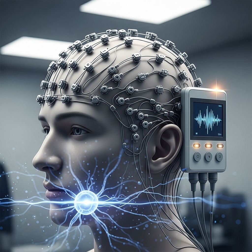
During an EEG, a technician places multiple electrodes on specific points of your head. These electrodes pick up electrical signals emitted by brain cells and send them to a computer, which records the data as wavy lines—also called brain waves—on a monitor or printout. Physicians analyze these patterns for anomalies that might indicate specific neurological disorders or events.
Why Is an EEG Performed?
EEGs are essential for diagnosing and monitoring a variety of conditions that affect brain health and function. Your doctor may order an EEG for several reasons:
- Diagnosing Epilepsy and other seizure disorders. EEGs are considered the gold standard for recognizing abnormal brain wave patterns associated with epileptic activity.
- Investigating Unexplained Seizures, fainting spells, or events that could be seizures.
- Determining Causes of Abnormal Brain Function, such as encephalopathies, that may present with confusion, abnormal movements, or altered mental status.
- Evaluating Head Injuries to assess potential brain trauma or monitor for seizures after a traumatic brain event.
- Identifying Sleep Disorders like narcolepsy or certain forms of insomnia.
- Assessing Brain Activity After Stroke, Infection, or Encephalitis when changes in consciousness or function occur.
- Determining Brain Death in patients in a coma, by confirming the absence or presence of electrical activity.
EEGs may also be used to monitor how effective treatment for seizures and other neurological disorders is working or to help plan brain surgery — for example, in patients with medication-resistant epilepsy who are surgical candidates.
Types of EEGs
The specific EEG used depends on your symptoms, the questions physicians need answered, and practical considerations related to duration and setting. The main types include:
- Routine EEG: This standard test usually takes 20-40 minutes with you lying still. You may be asked to perform specific tasks, such as deep breathing (hyperventilation) or looking at flashing lights (photic stimulation), to provoke possible changes in brain activity.
- Sleep EEG: In cases where routine tests are inconclusive, your doctor may request an EEG while you’re asleep. This can reveal sleep-related brain abnormalities that are otherwise hidden.
- Prolonged or Ambulatory EEG: For patients who rarely experience symptoms or when routine testing fails to capture abnormal activity, a continuous EEG monitoring system may be used. Small, portable EEG devices record brain activity over 24 to 72 hours, enabling you to go about most of your normal activities.
- Video EEG Monitoring: In certain cases, particularly in the diagnosis of complex seizure types, EEG recording is paired with video monitoring. This helps doctors correlate observed physical symptoms with electrical brain activity.
- Invasive EEG (Intracranial EEG): Rarely, for surgical planning or when non-invasive tests are inadequate, doctors may place electrodes directly on the surface or inside the brain through surgery. This is most commonly reserved for pre-surgical evaluation in epilepsy.
Preparing for an EEG
EEGs are safe and require little preparation, but some specific steps can help ensure the reliability of the test:
- Hair Preparation: Wash your hair the night before and do not use hair products, oils, or sprays; clean, product-free hair helps electrodes adhere and conduct signals.
- Fasting: Do not fast or restrict fluids unless specifically instructed to do so. Some EEGs require you to sleep less than usual, so follow your physician’s instructions about sleep deprivation if asked.
- Medication: Continue taking your usual medications unless told otherwise. Some drugs can affect EEG results, so bring a current list of all your medications (prescription and over-the-counter).
- Clothing: Wear comfortable clothing and avoid jewelry or accessories that may interfere with electrode placement.
- Other Instructions: Arrive on time. If you are having an ambulatory EEG, ask if you need to arrange for someone to drive you home.
How Is an EEG Performed?
Most EEGs are done in an outpatient clinic, hospital, or occasionally at the bedside in inpatient or intensive care settings. Here is what to expect:
- Preparation: You are asked to lie down or sit in a relaxed position. A trained technician will measure your head and mark electrode locations using a special template or cap. Your scalp will be cleaned to remove oils so electrodes can make good contact.
- Electrode Placement: Small electrodes, which look like flat metal discs, are applied to specific parts of your scalp using a sticky paste or gel. In some EEGs, an elastic cap with built-in electrodes is used.
- Recording: Once all electrodes are in place and connected to the EEG machine, recording begins. You do not feel the electrical impulses; the test is entirely painless, though the paste or gel may feel slightly cool.
- Provocation: You might be asked to perform deep breathing or exposed to flashing lights, which can trigger abnormal brain waves in certain conditions.
- Stillness: Movements, talking, or clenching your jaw can interfere with the readings. You will be asked to remain as still and relaxed as possible.
- Completion: When the test is finished, the electrodes are gently removed. Any remaining paste is wiped off, though you may wish to wash your hair after returning home.
Depending on the type of EEG, the test may last from 20–40 minutes (routine) up to several hours or days (prolonged/ambulatory EEGs).
What Does an EEG Detect?
EEG provides a way to assess many aspects of brain function and dysfunction. It’s an important tool for neurologists because it can:
- Detect abnormal electrical activity associated with seizures or epilepsy.
- Identify slowing, attenuation, or absence of electrical activity in coma or brain injury.
- Reveal patterns suggestive of tumors, stroke, encephalitis, or metabolic disorders affecting the brain.
- Assess effects of sedatives, anesthetics, or toxic exposures on the brain.
- Help diagnose sleep disorders by showing characteristic changes in brain wave patterns during sleep stages.
However, some brain conditions will not always show up on an EEG, or may require multiple or prolonged testing for abnormalities to be recorded.
Understanding EEG Results
After an EEG, a specially trained neurologist interprets the recorded brain wave patterns. Key points include:
- Normal EEGs show predictable patterns, varying with the patient’s age, level of alertness, and state of wakefulness or sleep.
- Abnormalities might include spikes, sharp waves, or unusual slowing. These findings can point to epilepsy, encephalopathy, or other issues.
- Transient findings (e.g., a single spike or brief irregularity) aren’t always cause for concern and may need to be interpreted in the context of your medical history.
- Further testing or monitoring might be needed if the EEG was inconclusive, incongruent with your symptoms, or conducted as part of longer-term diagnostic planning.
Risks and Safety Considerations
EEGs are non-invasive, safe, and painless. The test does not deliver any electrical current to your brain or body. The risks are minimal, but a few considerations include:
- Skin irritation: The adhesive, paste, or gel may cause mild skin irritation, especially for people with sensitive skin.
- Seizure risk: In rare cases, EEGs (especially those that use photic stimulation or hyperventilation) can trigger a seizure in individuals with seizure disorders. Medical personnel trained to handle seizures will always be present.
- Discomfort: Prolonged EEG monitoring may lead to temporary discomfort from wearing the electrodes or cap for extended periods.
There are no known long-term side effects or complications from the EEG procedure.
Advances and Innovations in EEG Technology
Technical and interpretive advances are continuously improving the diagnostic power of EEG:
- Quantitative EEG (qEEG): Computerized analysis of EEG data offers new ways to detect subtle changes, such as those found in early-stage brain injury or neurological disease. qEEG uses complex algorithms and pattern recognition to reveal abnormalities that may not be apparent on visual inspection alone.
- EEG Network Analysis: Innovations like network-based analysis examine how different brain regions interact and help distinguish conditions like epilepsy even when the EEG appears normal. Recent research, including tools like “EpiScalp,” can cut misdiagnoses by uncovering hidden patterns in routine EEGs, helping differentiate epilepsy from conditions that mimic seizures.
- EEG Combined with Imaging: Techniques that merge EEG data with MRI, CT, or PET brain scans can improve diagnostic accuracy and localize problems for treatment or surgery.
Limitations of EEG
While EEGs are invaluable for certain neurological diagnoses, they do have limitations:
- Cannot detect all brain problems: Some issues, including those deep within the brain, may not show up on scalp EEG.
- Normal results do not always rule out epilepsy or other neurological diseases. For some conditions, multiple or prolonged recordings may be necessary because abnormal electrical activity can be intermittent and easily missed during a brief session.
- Results depend on patient cooperation and absence of movement: Movement, muscle clenching, or talking during the test can create artifacts that are difficult to interpret.
Frequently Asked Questions (FAQs)
Q: Is an EEG painful or dangerous?
A: No. EEGs are completely non-invasive and painless. You may feel a cool sensation from the electrode gel or mild discomfort from sitting still, but there is no pain or electric shock involved.
Q: Will I have to stay overnight?
A: Most EEGs are done on an outpatient basis and take under an hour. Only prolonged or sleep EEGs may require overnight or multi-day monitoring.
Q: Can I eat or take my meds before the test?
A: Yes. Unless your doctor gives special instructions, eat as usual and continue your regular medications.
Q: Will an EEG show the exact cause of my symptoms?
A: An EEG can provide important information about brain activity and may support or clarify a diagnosis. However, it is always used alongside your medical history, other tests, and doctor evaluation—results are interpreted in the broader clinical context.
Q: What happens if my EEG is normal but I still have symptoms?
A: A normal EEG does not exclude all neurological problems. Some abnormalities may not occur during the recording, or may need longer or repeated monitoring to be captured.
Summary Table: EEG at a Glance
| Aspect | Details |
|---|---|
| Test Name | Electroencephalogram (EEG) |
| Purpose | Measures electrical brain activity to diagnose neurological disorders |
| Preparation | Wash hair, avoid hair products, follow doctor’s special instructions |
| Duration | 20–40 minutes (routine), up to several days (prolonged) |
| Risks | Minimal; rare risk of seizure in susceptible people |
| Recovery | Immediate; resume normal activities after the test |
| Limitations | May not always detect intermittent or deep-brain abnormalities |
References
- Johns Hopkins Medicine: Electroencephalogram (EEG)
- The Defeating Epilepsy Foundation
- BME Johns Hopkins News and Publications
- Annual Review of Biomedical Engineering – Advances in qEEG Analysis
References
- https://www.bme.jhu.edu/news-events/news/new-epilepsy-tool-could-cut-misdiagnoses-by-nearly-70-using-routine-eegs/
- https://pure.johnshopkins.edu/en/publications/advances-in-quantitative-electroencephalogram-analysis-methods-4
- https://www.defeatingepilepsy.org/medical-diagnostic-series/electroencephalogram-eeg/
- https://pmc.ncbi.nlm.nih.gov/articles/PMC9436243/
- https://pure.johnshopkins.edu/en/publications/diagnosing-epilepsy-with-normal-interictal-eeg-using-dynamic-netw
- https://pubmed.ncbi.nlm.nih.gov/8458997/
- https://jamanetwork.com/journals/jamaneurology/fullarticle/795101
Read full bio of Sneha Tete


