Diabetic Retinopathy & OCT: Modern Diagnosis and Management
An in-depth guide to how optical coherence tomography (OCT) helps diagnose and monitor diabetic retinopathy, with essential facts for patients.

Diabetic Retinopathy & Optical Coherence Tomography (OCT)
Diabetic retinopathy (DR) is a progressive eye disease caused by high blood sugar damaging the retinal blood vessels, affecting more than half of all people with diabetes. Optical coherence tomography (OCT) is a modern, noninvasive imaging technique eye doctors use to detect and monitor DR at its earliest stages, often before symptoms appear. This article explores how OCT works, its role in diagnosis and management, alternative detection methods, symptoms, medical help, and expert responses to frequently asked questions.
Why Regular Eye Exams Are Critical for Diabetes Management
People living with diabetes may not notice symptoms of diabetic retinopathy until the disease has advanced. Early damage typically goes undetected without specialized screening, which is why regular eye exams and diagnostic tests are essential for preserving vision.
- Prevention of vision loss: Early detection allows prompt treatment and monitoring.
- Silent progression: Retinopathy rarely causes noticeable symptoms in early stages.
- Routine screening: Eye doctors use advanced imaging and blood tests to check for underlying retinal changes.
What Is Diabetic Retinopathy?
Diabetic retinopathy develops when high blood glucose levels damage the retinal blood vessels. Over time, this leads to fluid leaks, swelling (macular edema), bleeding, and eventually scarring or retinal detachment. Untreated, it can result in partial or complete vision loss.
- Prevalence: Affects at least 50% of people with diabetes.
- Stages:
- Non-proliferative: Early phase with microaneurysms and minor swelling.
- Proliferative: Advanced phase marked by retinal bleeding and abnormal blood vessel growth.
Optical Coherence Tomography (OCT): Detailed Retinal Imaging
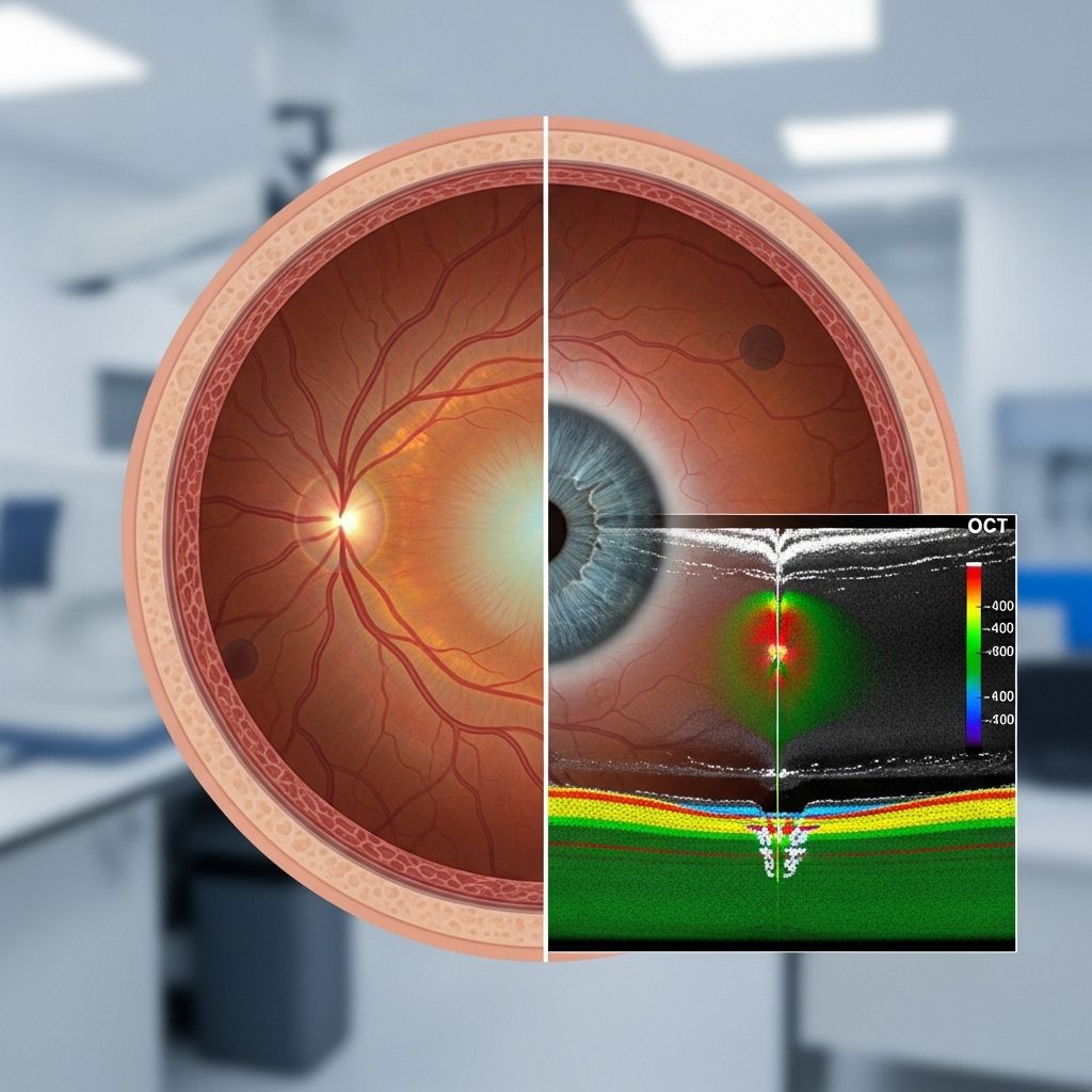
OCT is an advanced diagnostic test that uses near-infrared light waves to create high-resolution cross-sectional images of the retina’s layers, revealing structural changes invisible to standard eye exams.
| Feature | Description |
|---|---|
| Technique | Noninvasive use of light waves to scan and visualize retina |
| Output | 3D color or black-and-white images, showing retinal layers & thickness |
| Advantages | Immediacy, accuracy, comfortable procedure, detects early damage |
| Other Uses | Diagnosis of glaucoma, macular edema, age-related macular degeneration, vitreous traction |
How Does an OCT Scan Detect Diabetic Retinopathy?
OCT enables ophthalmologists to:
- Measure retinal thickness and identify swelling or fluid buildup.
- Visualize abnormal changes such as blood vessel leakage, hemorrhages, and microaneurysms.
- Monitor disease progression by comparing current images with previous results.
The rapid analysis possible with OCT means that doctors can discuss findings and adjust treatment during the same visit.
OCT’s Role in Managing Diabetic Retinopathy
OCT is not only invaluable for diagnosis—it is a primary tool to monitor DR progression.
- Routine monitoring: Regular OCT scans track changes over time, helping to assess treatment efficacy and detect new swelling or bleeding.
- Early intervention: Subtle findings prompt timely therapy, preventing further damage.
- Comparison of scans: Sequential imaging identifies minute changes, facilitating personalized care.
OCT Scan Procedure: What to Expect
This quick, painless procedure is performed in the eye doctor’s office. Here’s a step-by-step overview:
- Preparation: You’ll be seated in front of an OCT machine. Dilating eye drops may be applied to widen your pupils (may cause light sensitivity for a few hours).
- Imaging: The scan itself takes 5–10 minutes. You’ll be asked to focus on a target while the device captures images.
- Review: Results are available immediately, and your doctor will discuss findings and next steps.
Other Tests Used to Detect and Diagnose Diabetic Retinopathy
OCT is part of a comprehensive suite of tests. Depending on individual needs, ophthalmologists may recommend:
- Lab Tests:
- Fasting blood glucose: Normal is typically ≤ 110 mg/dL.
- Hemoglobin A1c (HbA1c): Indicates average blood glucose over past 2–3 months.
- Dilated Eye Exam:
- Pupils are dilated with drops, allowing a thorough retina examination.
- Detects retinopathy, cataracts, glaucoma, and other eye problems.
- May be repeated every 2–4 months if DR is diagnosed.
- OCT Angiography (OCT-A):
- Produces images of retinal blood vessels (unlike standard OCT, which shows structure).
- Important for measuring disease progression.
- Fluorescein Angiogram:
- Uses dye to visualize retinal blood vessels and leakage.
- Highly detailed images help pinpoint vascular changes.
- Funduscopic (Fundus) Exam:
- Direct imaging of retina for hemorrhages, vein/artery changes, microaneurysms.
- Ultrasonography (B Scan):
- Detects signs of retinal or vitreous detachment, especially helpful in late-stage DR.
Warning Signs & Symptoms of Diabetic Retinopathy
In early stages, diabetic retinopathy causes no symptoms. As the disease progresses, warning signs may include:
- Blurry vision
- Floaters (tiny spots or threads drifting in sight)
- Difficulty seeing at night
- Dark or empty areas in field of vision
- Sudden loss of vision
Because these symptoms often appear only after extensive damage, regular eye screening is critical for all people with diabetes.
When to Seek Medical Help
If you experience any changes in vision, it is crucial to contact your eye doctor promptly. Prompt detection and intervention is key to preserving sight. Routine eye exams and recommended imaging (including OCT) should be maintained even if you feel well or have no symptoms.
- Don’t wait for noticeable changes—early retinopathy may cause irreversible vision loss before being detected without screening.
- Annual checks are recommended for most adults with diabetes. More frequent visits may be advised based on disease stage and risk factors.
Frequently Asked Questions (FAQs)
What is OCT, and why is it important in diabetic retinopathy?
OCT stands for Optical Coherence Tomography, a cutting-edge imaging method producing cross-sectional, high-resolution images of the retina. It is crucial in diabetic retinopathy because it reveals minute changes in the retinal structure and thickness that can signal early-stage disease, permitting prompt intervention.
Is an OCT scan painful or risky?
No. The test is noninvasive, painless, and safe. You may experience brief discomfort from bright light or temporary sensitivity from dilating drops. Otherwise, there are no known serious risks with OCT.
How does an OCT scan differ from other imaging tests?
Unlike traditional fundus photography (which captures surface images) and angiography (which uses dyes to visualize blood vessels), OCT provides detailed, layered analysis of the retina’s structure, offering early detection and comprehensive monitoring.
Can diabetic retinopathy be cured?
Diabetic retinopathy cannot be reversed, but it can be managed. Early detection and treatment slow progression and preserve vision. Modern therapies—such as laser treatments, injections, or surgery—are highly effective if caught early.
How often should diabetic patients have OCT scans?
Frequency depends on disease status:
- No retinopathy: Annual eye exam, including OCT.
- Retinopathy present: OCT scans every 2–4 months or as your ophthalmologist recommends.
Summary
Diabetic retinopathy is a leading cause of vision impairment in people with diabetes. OCT scanning has transformed detection, monitoring, and management by providing detailed structural retinal images quickly and safely. Alongside other diagnostic tests and early routine eye exams, OCT is the cornerstone in fighting vision loss from diabetes. If you have diabetes, regular discussions with your ophthalmologist and scheduled checkups are essential to keeping your eyes healthy and your vision sharp.
References
- https://www.healthline.com/health/eye-health/diabetic-retinopathy-oct
- https://pmc.ncbi.nlm.nih.gov/articles/PMC12248572/
- https://pmc.ncbi.nlm.nih.gov/articles/PMC5723235/
- https://www.healthline.com/health/type-2-diabetes/retinopathy
- https://www.medicalnewstoday.com/articles/ozempic-semaglutide-may-help-protect-against-diabetes-related-blindness-retinopathy
- https://www.mayoclinic.org/diseases-conditions/diabetic-retinopathy/symptoms-causes/syc-20371611
- https://www.ccjm.org/content/91/8/503
Read full bio of medha deb


