Cystography: Understanding Bladder Imaging, Procedures, and What to Expect
A comprehensive guide to cystography—bladder imaging procedures, reasons, risks, safety, preparation, and recovery.

Cystography is a diagnostic imaging procedure designed to evaluate the structure and function of the bladder and related portions of the urinary tract. Often using X-ray technology and special contrast dyes, cystography provides invaluable insights for clinicians investigating causes of urinary symptoms, recurring infections, trauma, and congenital abnormalities.
What Is Cystography?
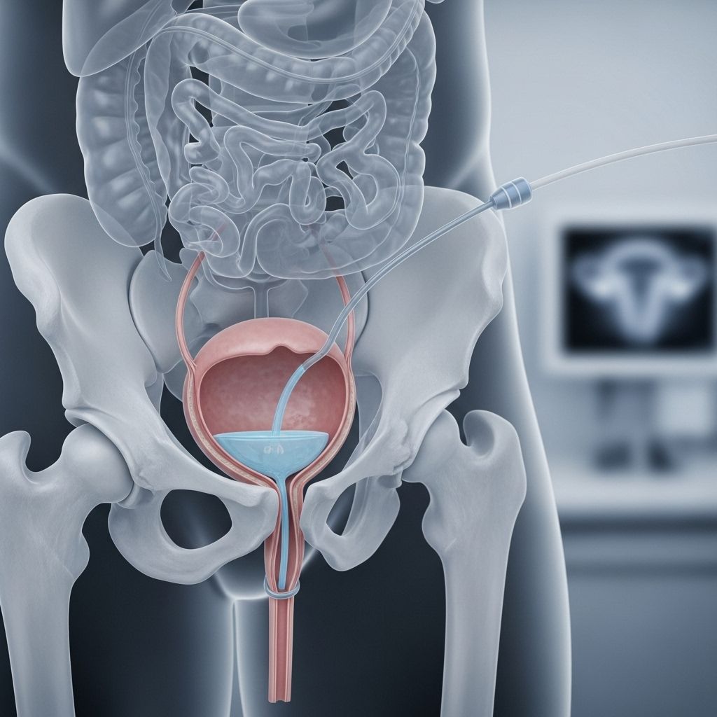
Cystography is an imaging test that visualizes the bladder using X-rays complemented by a contrast dye introduced into the bladder via a catheter. This procedure allows health care providers to detect anatomical and functional abnormalities in the bladder, assess bladder injuries, track infections, and evaluate issues relating to urination. There are several variations of cystography, including retrograde cystography, CT cystography, and voiding cystourethrography.
- Standard cystography: X-ray pictures are taken while the bladder is filled with contrast dye.
- Fluoroscopic cystography: Provides real-time moving images of the bladder during filling and voiding.
- Voiding cystourethrography (VCUG): Captures images as the patient urinates to assess urinary flow and reflux.
- CT cystography: Utilizes advanced CT imaging for detailed investigation, often used after trauma.
- Radionuclide cystogram: Uses radioactive material to check bladder function, often for specialized cases.
Why Is Cystography Performed?
Physicians recommend cystography for several reasons—typically when other tests fail to fully explain urinary symptoms or when imaging is necessary to guide further management.
- Blood in urine (Hematuria): To find structural sources of bleeding, including masses or stones.
- Recurrent urinary tract infections (UTIs): To detect anatomical defects or reflux that predispose to infection.
- Suspected bladder injury: Following trauma, particularly pelvic fractures, to identify ruptures or leaks.
- Dysuria or voiding difficulty: For patients with symptoms like painful urination, urgency, or inability to empty the bladder fully.
- Evaluation of vesicoureteral reflux (VUR): When urine flows backward from the bladder to the kidneys, risking infection and kidney damage.
- Assessment of anatomical defects: Congenital or acquired bladder problems, diverticula, tumors, or fistulas.
Cystography is sometimes employed after surgery to ensure the bladder is healing properly or to check for leakages.
Types of Cystography
| Test Type | Description | Common Uses |
|---|---|---|
| Plain X-ray cystography | Contrast dye fills the bladder, followed by X-ray images | Suspected bladder injury, tumors, reflux |
| Fluoroscopic cystography | Real-time imaging during filling and emptying phases | Bladder function assessment, voiding dysfunction |
| CT cystography | CT scans after contrast introduction | Detect complex injuries post-trauma |
| Radionuclide cystogram | Radioactive tracer with nuclear scanner | Vesicoureteral reflux in children, functional exams |
How to Prepare for Cystography
Preparation for cystography is generally straightforward, but may vary slightly by test type and patient age:
- A signed informed consent is mandatory for all patients.
- Empty the bladder before commencing the test unless instructed otherwise.
- You may be asked about allergies, especially to iodine or contrast dyes.
- Discuss all medications and recent illnesses with your doctor.
- Remove jewelry and metallic objects, which can interfere with imaging.
- Wear a hospital gown provided by the facility.
Fasting is rarely necessary. For children, supportive explanation beforehand can help reduce anxiety related to catheterization and imaging.
What Happens During Cystography?
Cystography is typically performed in a radiology department, using a combination of sterile technique, mild sedation (in select cases), and local numbing for maximum comfort.
- Set-up: The patient lies on an X-ray table. The genital area is cleaned with antiseptic solution.
- Catherization: A thin, flexible catheter is inserted through the urethra into the bladder. Lubricant and numbing medicines are often used to minimize discomfort.
- Filling the bladder: The bladder is gently filled with a contrast dye—or radioactive liquid, in the case of radionuclide cystograms—through the catheter until full, or until the patient indicates fullness or discomfort.
- Imaging: As the bladder fills, a series of X-ray images (or, for certain tests, live fluoroscopic images or CT scans) are taken in different patient positions to view all aspects of the bladder wall and other structures.
- Voiding phase (if indicated): For a voiding cystourethrogram (VCUG), images are captured as the patient begins to urinate; this can reveal reflux or functional bladder issues.
- Completion: After imaging, the catheter is carefully removed and a final X-ray may be taken post-voiding to assess for residual urine or abnormal leaks.
The entire process usually takes 30 to 60 minutes, depending on the complexity of the case and the test variation.
What Will I Feel During Cystography?
- Insertion of the catheter may cause a sensation of pressure or mild discomfort.
- As the bladder fills with contrast, patients often feel a strong urge to urinate and fullness in the pelvis.
- Some temporary burning or discomfort may be experienced when urinating post-exam.
- Pain beyond mild soreness or fever should prompt a call to your provider.
Young children may require gentle restraint or distraction during the exam. Adults and most children tolerate cystography well with local anesthesia and support.
Risks and Complications of Cystography
While cystography is generally safe, as with any invasive or diagnostic procedure, some risks exist:
- Possible urinary tract infection (UTI)—due to catheterization.
- Rarely, allergic reaction to contrast dye (rash, swelling, difficulty breathing).
- Slight bleeding in urine immediately after the test.
- Temporary discomfort or burning during urination.
- Radiation exposure—generally low and considered safe, but minimized especially for children.
- In very rare cases, injury to the bladder or urethra from the catheter.
Discuss your allergies, health conditions, and any possible pregnancy with your provider to ensure appropriate precautions. For minors or pregnant individuals, alternative imaging such as ultrasound or minimizing radiation may be considered.
What Happens After Cystography?
Most patients can return home and resume regular activities immediately after cystography unless sedated or advised otherwise.
- Drink plenty of fluids to help flush the contrast dye and minimize infection risk.
- Temporary discomfort or burning with urination is common; this should resolve within a day.
- Monitor for signs of infection: fever, chills, persistent pain, heavy bleeding, or difficulty urinating.
- In children, additional reassurance may be necessary if discomfort persists.
Healthcare providers review the cystogram images and discuss the findings with you, which may guide further evaluation or treatment.
Results of Cystography
Your provider interprets the images to assess for:
- Bladder wall abnormalities: tumors, diverticula, polyps, or irregular thickening.
- Leakage of contrast out of the bladder—suggestive of trauma or surgical complication.
- Vesicoureteral reflux—visualized as contrast moving upward into the ureters and kidneys.
- Residual contrast after voiding—may indicate incomplete bladder emptying, an obstruction, or loss of muscle function.
- Foreign bodies or anatomical variants.
Further management will depend on test results, symptoms, and additional diagnostic findings. Your provider will explain any abnormalities and outline next steps.
Frequently Asked Questions (FAQs)
What is the difference between cystography and cystoscopy?
Cystography is an X-ray imaging test using contrast dye to visualize the bladder, whereas cystoscopy is an endoscopic procedure in which a camera is inserted directly through the urethra for visual inspection and sometimes biopsy of the bladder lining.
Is cystography painful?
Cystography may cause mild, brief discomfort during catheter insertion and when the bladder is being filled; most patients tolerate the procedure well, and numbing agents are often used. Discomfort should subside soon after the test.
Can I eat or drink before the procedure?
Fasting is not usually required for cystography. However, follow your health care provider’s specific instructions, particularly if sedation will be used.
How long do results take?
Preliminary results are often available shortly after the test. A radiologist or urologist reviews the images in detail and discusses the findings with you during a follow-up appointment.
Are there alternatives to cystography?
Depending on the clinical question, ultrasound or MRI may sometimes be used to evaluate bladder anatomy and function without radiation or contrast dye. These alternatives may not be as detailed for certain pathologies.
Is cystography safe for children?
Cystography is commonly performed in children suspected of vesicoureteral reflux or recurrent UTIs. Steps are taken to ensure safety and minimize radiation exposure. The benefits of accurate diagnosis usually outweigh the small risks.
How do I care for myself after the procedure?
Hydrate well, note any lasting pain, fever, or abnormal urine, and follow up with your doctor regarding results. Most people experience few or no complications.
Summary
Cystography is a valuable medical imaging tool for diagnosing a wide array of bladder and lower urinary tract issues. It is generally safe, easy to perform, and highly informative for both acute and chronic conditions. If your provider recommends cystography, being informed will help you feel comfortable and prepared throughout the process.
References
- https://www.urmc.rochester.edu/encyclopedia/Content?contentTypeID=92&ContentID=P07771
- https://www.mountsinai.org/health-library/tests/radionuclide-cystogram
- https://healthlibrary.inova.org/BreatheEasy/92,P07704
- https://www.ucsfbenioffchildrens.org/medical-tests/retrograde-cystography
- https://www.mayoclinic.org/tests-procedures/cystoscopy/about/pac-20393694
- https://my.clevelandclinic.org/health/diagnostics/16553-cystoscopy
- https://www.mskcc.org/cancer-care/patient-education/about-your-cystogram
- https://ufhealth.org/conditions-and-treatments/cystoscopy
Read full bio of medha deb


