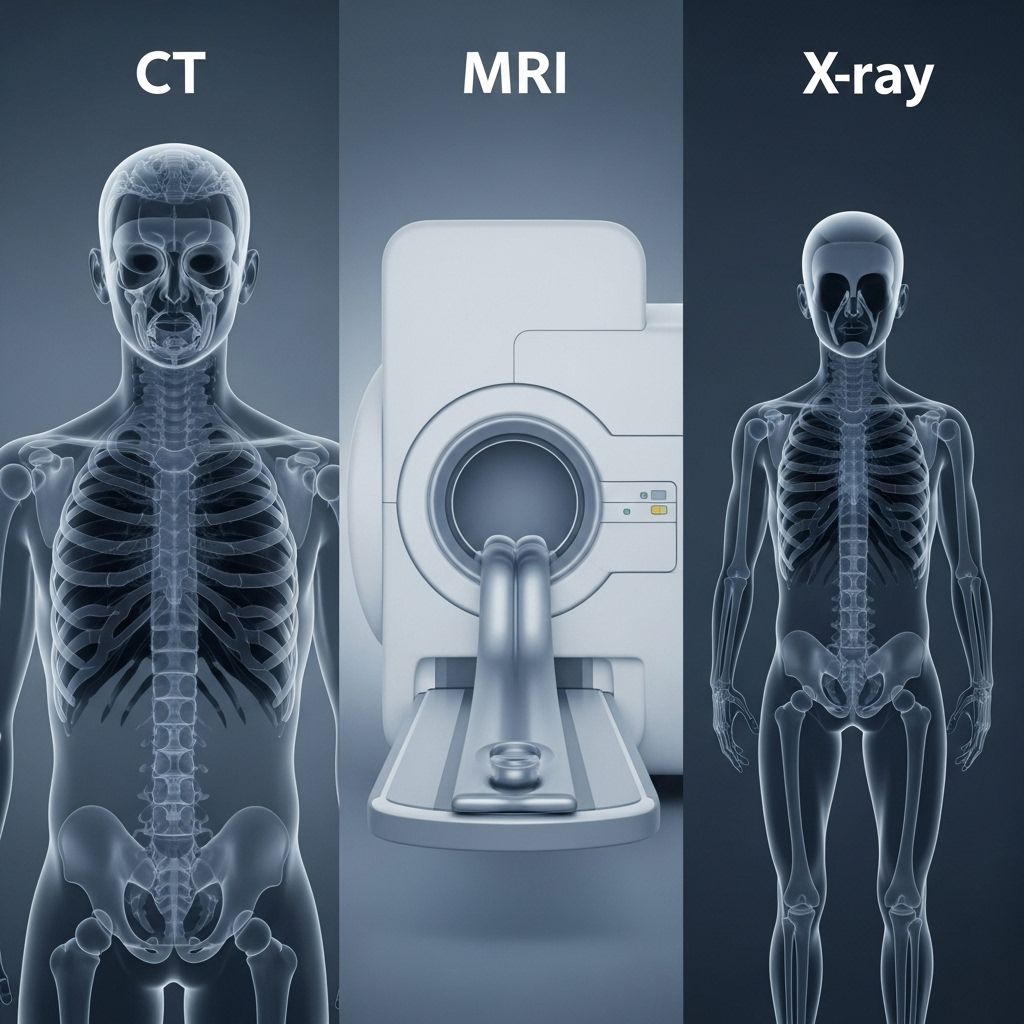
CT vs. MRI vs. X-Ray: Key Differences in Medical Imaging
Understand the differences, uses, and risks of CT scans, MRIs, and X-rays to make informed choices about diagnostic imaging.

CT vs. MRI vs. X-Ray: Understanding Diagnostic Imaging
Medical imaging has revolutionized modern healthcare, allowing doctors to look inside the body without surgery. CT scans, MRIs, and X-rays are three of the most common imaging tests, each with unique applications, benefits, and limitations. Understanding the differences among these technologies can help patients and families participate in health decisions and feel prepared for diagnostic procedures.
Table of Contents
- Introduction to Medical Imaging
- What is an X-Ray?
- What is a CT Scan?
- What is an MRI?
- Similarities Among X-Ray, CT, and MRI
- Key Differences: CT vs. MRI vs. X-Ray
- Choosing the Right Imaging Test
- Risks and Safety Considerations
- Frequently Asked Questions
Introduction to Medical Imaging
Medical imaging refers to techniques that create visual representations of the interior of the body. These images are used to diagnose, monitor, and sometimes treat diseases. The most common imaging tests are:
- X-rays: First widespread imaging technology, in use for over a century.
- CT (Computed Tomography) scans: Advanced imaging using multiple X-ray images from different angles to create 3D pictures.
- MRI (Magnetic Resonance Imaging): Uses strong magnets and radio waves to produce highly detailed images of tissues and organs.
What is an X-Ray?
An X-ray is a quick, noninvasive test that helps doctors diagnose conditions by viewing structures inside the body, especially bones.
How X-Rays Work
X-ray machines pass a controlled burst of X-ray radiation through the body. Dense structures like bones absorb more radiation and appear white on the X-ray image, while softer tissues allow more radiation to pass through and show up in shades of gray.
Common Uses for X-Rays
- Bone fractures and dislocations
- Detecting tooth problems in dental exams
- Finding pneumonia, lung infection, or lung cancer in the chest
- Spotting some types of tumors
What to Expect During an X-Ray
- The test is quick—usually less than 15 minutes.
- You may be asked to stand, sit, or lie down depending on the body part being imaged.
- Lead shields may be used to protect other parts of the body from unnecessary exposure.
- You usually will not feel anything during the imaging process.
What is a CT Scan?
A CT scan, or Computed Tomography, uses X-ray technology to capture multiple images—or “slices”—of the body from various angles. These slices are combined by a computer to create detailed 3D images, offering more information than standard X-rays.
How CT Scans Work
A CT scanner is a large, donut-shaped machine. The patient lies on a narrow table that slides through the center of the scanner. X-ray beams rotate around the body, collecting cross-sectional images that are reconstructed by a computer.
Common Uses for CT Scans
- Identifying head injuries, such as bleeding or stroke
- Evaluating bone fractures not visible on X-rays
- Detecting and monitoring tumors or masses
- Diagnosing and assessing internal organ damage (lungs, liver, kidneys)
- Detecting blood clots or bleeding in the body
- Guiding biopsies or certain minimally invasive procedures
What to Expect During a CT Scan
- Most CT scans take about 10–30 minutes.
- You may be asked to hold your breath to limit motion.
- Contrast dye (oral or intravenous) may be used to enhance images.
- The test is painless, but the scanner can be noisy.
What is an MRI?
A Magnetic Resonance Imaging (MRI) scan uses strong magnets and radio waves instead of X-rays to generate detailed images of the organs and tissues within the body.
How MRIs Work
During an MRI, the patient lies inside a tube-like machine that generates a powerful magnetic field. Radiofrequency pulses bounce off water and fat molecules in the body, producing signals detected by the scanner and used to build 3D images.
Common Uses for MRIs
- Examining the brain and spinal cord for diseases and injuries
- Imaging muscles, ligaments, and tendons (sports injuries, tears, or strains)
- Diagnosing some types of tumors and cysts
- Assessing breast tissue or certain abdominal organs
- Evaluating heart and blood vessel abnormalities
What to Expect During an MRI
- MRI scans usually take 30 to 60 minutes or longer.
- The scanner can be noisy, producing clicking and thumping sounds.
- You’ll be asked not to move during the scan; earplugs or headphones may be provided.
- Some people may feel claustrophobic in the narrow tube.
- Contrast agents may be administered intravenously for some studies.
Similarities Among X-Ray, CT, and MRI
- All are noninvasive diagnostic imaging tests that help doctors visualize the interior of the body.
- Patients need to remain still to obtain clear images.
- Tests are generally painless, though some may feel uncomfortable due to positioning or confinement.
- Results from these imaging tests can guide further treatment decisions or track disease progress.
Key Differences: CT vs. MRI vs. X-Ray
| Feature | X-Ray | CT Scan | MRI |
|---|---|---|---|
| How It Works | X-ray radiation | Multiple X-ray images + computer reconstruction | Magnetic field and radio waves |
| Uses Radiation? | Yes | Yes (higher dose) | No |
| Typical Imaging Time | Few minutes | 10–30 minutes | 30–60 minutes or more |
| Image Detail | Good for bones, some soft tissue | Detailed images of bones, organs, soft tissue | Highly detailed, especially soft tissues |
| Common Uses | Broken bones, chest infections, dental | Injuries, tumors, internal organ issues, trauma | Brain, spine, joints, muscles, some tumors |
| Exposure Limitations | Not recommended for pregnancy (unless necessary) | Higher radiation exposure than X-ray | Unsafe for patients with certain implants, metal in body |
| Cost | Least expensive | Moderate | Most expensive |
| Contrast Agents | Rarely | Often (iodine-based) | Sometimes (gadolinium-based) |
Choosing the Right Imaging Test
Doctors select the most appropriate imaging test based on the suspected diagnosis, the body part involved, patient history, and safety considerations. Sometimes, more than one type of imaging is necessary for a comprehensive diagnosis.
- X-ray is often the first imaging test for suspected bone injuries or chest infections.
- CT scan is used when more detail is needed, such as in trauma, suspected internal bleeding, or certain cancers.
- MRI is preferred for neurological, musculoskeletal, or soft tissue concerns.
Risks and Safety Considerations
X-Ray and CT Scan Risks
- Both tests expose the body to ionizing radiation.
- Exposure levels are usually low and considered safe for most patients, but risks can accumulate with repeated exposure.
- CT scans deliver higher doses of radiation than standard X-rays.
- Contrast agents used in some CT scans may cause allergic reactions or affect kidney function in rare cases.
MRI Risks
- No ionizing radiation; considered safe for most people.
- Not suitable for patients with certain metal implants (e.g., pacemakers, aneurysm clips, some cochlear implants), as the magnetic field can pose risks.
- Some patients may experience anxiety or claustrophobia inside the machine.
- Contrast agents are generally safe but can cause rare allergic reactions.
Frequently Asked Questions (FAQs)
Q: Do X-rays, CT scans, or MRIs hurt?
A: These imaging tests are painless. You may experience minor discomfort from staying still, positioning, or, in the case of CT or MRI, lying on a table inside the scanning machine. Contrast dyes (if used) may cause mild, temporary side effects.
Q: Are imaging tests safe for children?
A: Medical imaging is widely used for children. Doctors tailor doses, minimize exposure, and typically avoid unnecessary scans, especially those involving radiation, to balance diagnostic needs with safety.
Q: How should I prepare for an imaging test?
A: Preparation varies by test. You may be asked to change into a gown or remove jewelry. For MRIs, inform your doctor of any implants or metal objects. You may need to avoid eating for a few hours beforehand if contrast dye is used.
Q: What if I am pregnant?
A: Always notify your healthcare provider if you are or might be pregnant. MRIs are typically considered safe during pregnancy, especially after the first trimester. X-rays and CT scans may be used only if absolutely necessary and with appropriate precautions.
Q: What happens after the imaging test?
A: A radiologist will review the images and send a report to your referring doctor, who will discuss the findings with you and recommend next steps if needed.
Key Takeaways
- X-rays are fast, affordable, and best for imaging bones and some chest or dental issues.
- CT scans provide detailed, cross-sectional images suitable for trauma, complex bone fractures, and certain cancers, but deliver higher radiation doses.
- MRIs offer exquisite soft-tissue detail for brain, spine, and muscle imaging without radiation but are slower and less suitable for patients with metal implants.
- Doctors consider medical history, condition, and risk factors when selecting an imaging modality.
Always consult your healthcare provider to understand the benefits and risks of any diagnostic test and to ensure the most appropriate imaging for your individual situation.
References
- http://www.wth.org/blog/ct-scans-mris-and-x-rays-oh-my-making-sense-of-imaging/
- https://www.envrad.com/difference-between-x-ray-ct-scan-and-mri/
- https://citywideradiology.com/mri-vs-ct-scan-vs-x-ray/
- https://upmc.ie/blog/radiology/know-the-difference-between-an-x-ray-ct-and-mri-scan
- https://orthoinfo.aaos.org/en/treatment/x-rays-ct-scans-and-mris/
- https://northcentralsurgical.com/whats-the-difference-between-an-x-ray-ct-scan-and-mri/
- https://www.prairie-ortho.com/blog/difference-between-x-rays-cts-and-mris/?bp=30476
- https://www.mskcc.org/news/ct-vs-mri-what-s-difference-and-how-do-doctors-choose-which-imaging-method-use
Read full bio of Sneha Tete


