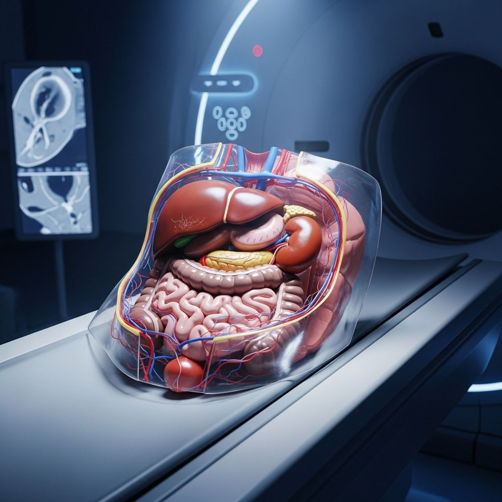
Computed Tomography (CT or CAT Scan) of the Abdomen
A comprehensive guide to abdominal CT scans: purpose, preparation, procedure, risks, and results for better healthcare decisions.

Abdominal computed tomography (CT) scans, also known as CAT scans, are a cornerstone of modern medical imaging. They offer rapid, detailed cross-sectional images of the internal organs, vessels, and structures within the abdomen. With advances in technology, CT imaging provides invaluable information for diagnostic, therapeutic, and surgical planning purposes. This guide explains what an abdominal CT scan is, why it is used, how you should prepare for it, the procedure itself, its potential risks, and what the results may mean.
What is an Abdominal CT Scan?
Computed Tomography (CT) involves the use of X-rays and computer processing to produce cross-sectional images (slices) of the abdomen. These images allow physicians to examine the various organs, blood vessels, and structures in much greater detail than standard X-rays. The scanner rotates an X-ray tube around the body, collecting images that are reconstructed into 2D and, when necessary, 3D representations.
- Also called a CAT scan (Computerized Axial Tomography).
- Produces detailed images of abdominal organs, vessels, bones, and soft tissue.
- Often performed with contrast agents (oral or intravenous) to enhance visibility of certain structures or abnormalities.
Why is a CT Scan of the Abdomen Performed?
- To detect the cause of abdominal pain, swelling, or discomfort.
- To identify the source of abnormal blood test results associated with kidney or liver function.
- To investigate unexplained fever or infections.
- To evaluate tumors, masses, cysts, or possible cancers (such as those affecting the liver, pancreas, kidneys, or bowel).
- To diagnose or monitor conditions such as hernia, kidney stones, appendicitis, bowel obstruction, inflammatory bowel disease, pancreatitis, and abdominal injuries.
- To help guide or plan for biopsies, minimally invasive procedures, or surgeries.
- To assess progress after surgical interventions or ongoing treatments, and to monitor for the spread of cancers originating elsewhere in the body.
Common Conditions Evaluated by Abdominal CT
- Renal and ureteric cancers
- Colon and liver cancers
- Lymphoma, melanoma, and ovarian cancer
- Pheochromocytoma, and other endocrine tumors
- Kidney infection, stones, and obstructions
- Hernias, acute cholecystitis (gallbladder inflammation), liver disease, pancreatitis
- Hydronephrosis (kidney swelling)
- Acute abdominal aortic aneurysm
- Abscesses or collections of infected fluid
- Inflammatory bowel conditions (Crohn’s disease, ulcerative colitis)
How Does a CT Scanner Work?
The CT scanner is a large, doughnut-shaped machine. The patient lies on a motorized table that moves slowly through the opening of the scanner (called the gantry). As the table advances, an X-ray tube rotates around the body, capturing images from multiple angles. These X-ray images are processed by a computer to reconstruct cross-sectional images—often less than a centimeter thick—that provide highly detailed views of the internal organs.
- Contrast Materials: Contrast agents (containing iodine or barium) may be administered orally, intravenously, or both to highlight blood vessels, bowel, or specific organs.
- Image Reconstruction: The software can combine slices to form 3D images, aiding analysis and surgical planning.
- Hounsfield Scale: Different tissues display unique values (air: -1000, water: 0, bone: +400 to +2000), helping to differentiate structures and pathologies.
How Should You Prepare for an Abdominal CT Scan?
Proper preparation ensures the highest quality images and helps reduce potential risks. Follow all instructions provided by your healthcare team.
- Fasting: You may need to avoid eating and drinking for several hours (often 2–6) before the examination, especially if contrast is to be used.
- Medication and Allergy Disclosure: Discuss any medications, allergies (especially to iodine, contrast agents, shellfish, or latex), recent illnesses, or chronic conditions with your physician or technologist.
- Metal and Clothing: Remove jewelry, piercings, and metallic objects. You may be asked to change into a hospital gown for comfort and image clarity.
- Pregnancy: Inform the technologist if you are—or might be—pregnant. CT scans involve radiation exposure, so alternative imaging may be selected for pregnant persons unless the benefit outweighs the risk.
- Pre-Scan Medications: If you have a history of allergic reactions to contrast media, premedication (such as steroids or antihistamines) may be prescribed to minimize the chance of a reaction.
What Happens During the CT Scan Procedure?
The abdominal CT is typically performed in a radiology or imaging department. The procedure is quick, noninvasive, and usually takes less than 30 minutes.
- Positioning: You will lie on your back (sometimes side or stomach), with arms overhead, on a narrow sliding table.
- Contrast Administration: If oral contrast is needed, you may be asked to drink a special liquid before the scan. If intravenous (IV) contrast is required, a small needle will be placed in your arm for the injection.
- Scanning: The table moves you into the scanner, and the X-ray tube rotates around the abdomen. You may hear quiet whirring or clicking sounds.
- Instructions: At various points, you might be instructed to hold your breath for a few seconds. Lying still is essential for clear images.
- Completion: Once the required images are captured, the IV (if used) is removed, and you can dress and leave the imaging suite.
What Will I Experience During and After the Procedure?
Most patients find the scan to be relatively comfortable. However, some sensations may occur, particularly if contrast agents are administered:
- Positioning: Lying on a hard table may cause mild discomfort.
- IV Contrast Sensations: Common experiences include a warm flush, slight burning at the injection site, or a metallic taste. These sensations typically fade in seconds.
- Reactions: Minor side effects from contrast, such as itching or mild rash, are possible. Severe allergic reactions are rare but require immediate medical attention.
- After the Scan: If contrast was used, you may be advised to drink extra fluids to help flush it from your body. Most people resume normal activities right away.
Who Interprets the Results and How Do You Get Them?
A board-certified radiologist will review the images, looking for abnormal findings. The radiologist generates a comprehensive report for your referring physician, who will discuss results and any necessary follow-up or treatment. If the findings are urgent or unexpected, results may be communicated more quickly.
Risks of an Abdominal CT Scan
- Radiation Exposure: CT scans use more radiation than standard X-rays. While the risk is small compared to the clinical benefit, multiple scans and high doses over time can increase cancer risk, particularly in children and pregnant individuals.
- Contrast Allergy or Kidney Effects: Allergic reactions to contrast agents are possible—ranging from mild to severe. Rarely, contrast can worsen existing kidney disease, especially in at-risk or dehydrated patients.
- Complications from Contrast: Nausea, headache, and rarely more serious effects such as very low blood pressure or anaphylaxis may occur. Oral contrast may cause mild gastrointestinal upset or rarely, obstruction.
- Pregnancy Considerations: Unnecessary exposure to radiation in pregnancy should be avoided when possible. Always notify your healthcare provider if you are (or might be) pregnant.
Benefits of Abdominal CT Scans
- Fast and Detailed: Provides high-resolution images rapidly, crucial in emergencies and acute care.
- Guides Treatment: Informs decision-making for surgery, biopsy, cancer therapy, and trauma management.
- Noninvasive and Painless: No incisions, no recovery time.
- Comprehensive Views: Simultaneously images multiple organs and systems.
What Do Abnormal Results Mean?
Interpretation of abnormal findings depends on the clinical question and the structures involved. Some common abnormal findings include:
- Tumors, masses, or suspicious lesions
- Evidence of internal bleeding or fluid collections
- Signs of infection, abscess, or inflammation
- Structural anomalies (e.g., hernias, aneurysms, obstructions)
- Kidney stones or evidence of organ injury
| Condition | Imaging Appearance |
|---|---|
| Appendicitis | Enlarged appendix, surrounding fat stranding |
| Kidney stones | Bright, dense calculi in urinary tract |
| Liver lesions | Solid or cystic masses, may enhance with contrast |
| Abdominal aneurysm | Enlarged aorta, sometimes with leak or rupture |
| Pancreatitis | Swollen, inflamed pancreas, possible fluid collections |
| Tumors | Irregular masses, often with necrosis or abnormal vasculature |
Limitations of Abdominal CT Scans
- Not Suitable for All Patients: Some patients, such as those with contrast allergies or severe kidney disease, may not be candidates for contrast-enhanced scans.
- Radiation Use: While individual scan risk is minimal, cumulative exposure should be minimized whenever possible.
- Soft Tissue Resolution: CT scans may not always differentiate among certain soft tissue abnormalities; MRI or ultrasound may be needed.
- Incidental Findings: Non-specific or unrelated abnormalities may be discovered, requiring further testing for clarification.
Alternative or Complementary Imaging Tests
- Ultrasound: Useful for some organ evaluation (e.g., gallstones, early pregnancy), especially suitable for children and pregnant individuals.
- MRI (Magnetic Resonance Imaging): Excellent for soft tissue contrast and doesn’t use ionizing radiation, but may not be as fast or widely available as CT.
- Plain X-ray: Sometimes used for preliminary evaluation, but much less detailed than CT.
Frequently Asked Questions (FAQs)
Q: How long does an abdominal CT scan take?
A: The entire process generally takes less than 30 minutes, with active scanning lasting only a few minutes. Extra time may be needed if contrast is administered.
Q: Is the scan painful?
A: The scan itself is painless. Temporary discomfort may result from lying on the table or from the injection of contrast material.
Q: Can I eat or drink before my scan?
A: You may be advised not to eat or drink for several hours before your scan, especially if contrast will be used. Follow your healthcare provider’s instructions closely.
Q: Are there risks of radiation exposure?
A: CT scans use ionizing radiation. While the clinical benefit typically outweighs the risk, unnecessary or repeated scans should be avoided, particularly in children and pregnant individuals.
Q: What if I am pregnant?
A: Always inform your provider if you are or could be pregnant. Alternatives to CT should be considered when possible to avoid exposing the fetus to radiation.
Q: Can I drive home after the scan?
A: Yes, you can usually resume normal activities immediately after your CT scan unless advised otherwise, particularly if you haven’t received any sedative medication.
Q: What should I do about contrast allergies?
A: If you have a known contrast allergy, notify your provider. Premedication and alternative imaging strategies are often available.
Summary
Abdominal CT scans are highly effective diagnostic tools used to evaluate a range of disorders affecting the organs and vessels of the abdomen. Used appropriately, they provide rapid, precise information that can guide urgent and non-urgent care. Always share medical history, allergies, and medication details with your healthcare providers to optimize safety and results.
References
- https://www.mountsinai.org/health-library/tests/abdominal-ct-scan
- https://www.radiologyinfo.org/en/info/abdominct
- https://www.ncbi.nlm.nih.gov/books/NBK567796/
- https://health.uconn.edu/radiology/wp-content/uploads/sites/195/2020/01/Radiology-CTscan.pdf
- https://my.clevelandclinic.org/health/diagnostics/4808-ct-computed-tomography-scan
- https://www.mayoclinic.org/tests-procedures/ct-scan/about/pac-20393675
- https://www.cedars-sinai.org/health-library/tests-and-procedures/c/ct-scan-of-the-abdomen.html
Read full bio of Sneha Tete


