Caliectasis: Symptoms, Causes, Diagnosis, and Treatment
Learn about caliectasis: a condition marked by swollen kidney calyces, its causes, symptoms, diagnosis, treatment, prevention, and potential complications.

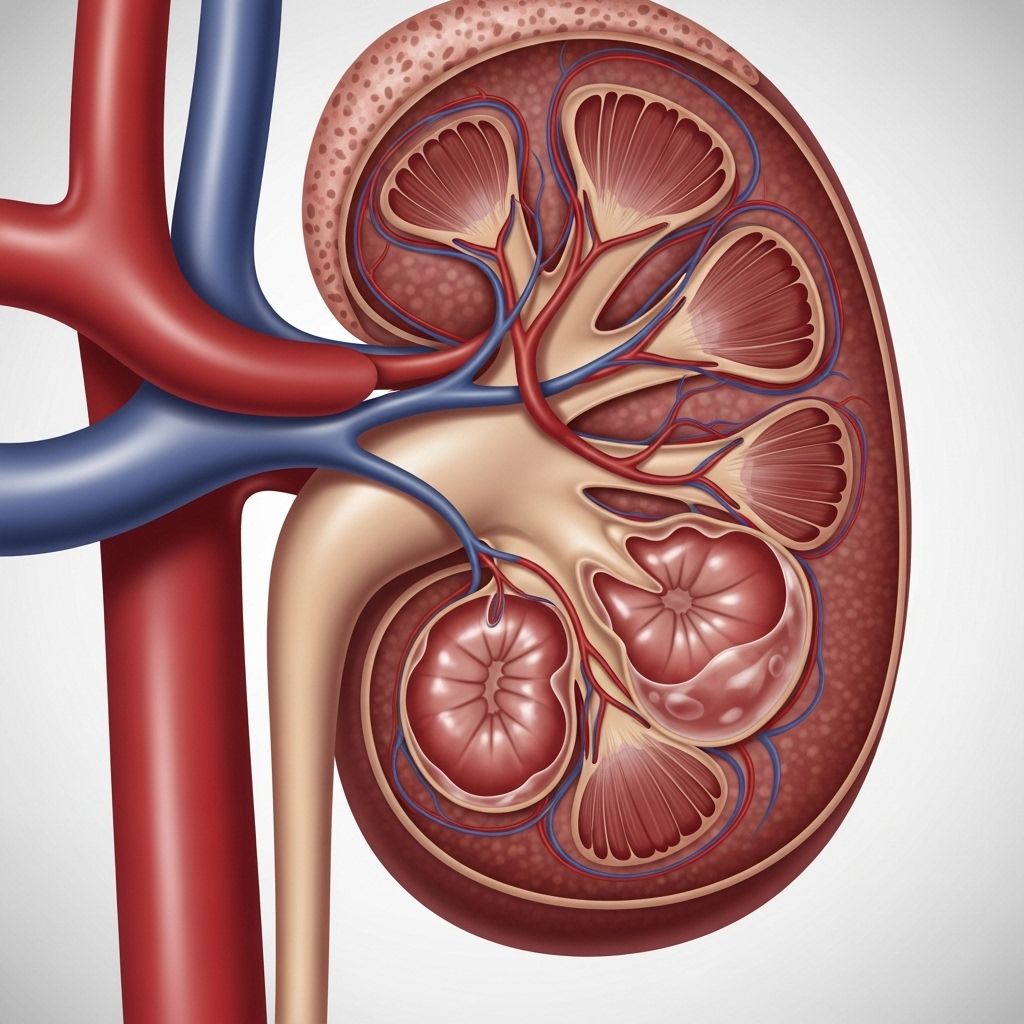
Caliectasis is a medical condition affecting the calyces within the kidneys. Calyces are cup-like urine-collecting structures—each kidney has 6 to 10—located near the outer edges of the organ. With caliectasis, these calyces become dilated and swollen due to fluid accumulation, commonly triggered by another kidney-related disorder. Early diagnosis and targeted treatment are essential for protecting kidney function and preventing complications.
What Is Caliectasis?
Caliectasis refers to the dilation or enlargement of the calyces inside one or both kidneys. The primary role of calyces is collecting urine formed in the kidney before it passes into the renal pelvis and then to the ureter and bladder. Caliectasis itself is rarely a standalone illness; it is typically a sign of an underlying issue causing an interruption or disturbance in normal urine flow or kidney function.
Diagnosing caliectasis usually requires radiological imaging—most people only discover it while being examined for other kidney-related symptoms or diseases.
Symptoms of Caliectasis
Caliectasis does not produce symptoms directly. Instead, symptoms stem from the underlying condition that disrupts urine flow or damages kidney structures. Common indirect symptoms include:
- Blood in urine (hematuria)
- Abdominal pain or tenderness
- Lower back pain
- Trouble urinating
- Increased urge to urinate or urinary frequency
- Difficulty urinating
- Pus in urine (pyuria)
- Foul-smelling urine
If you experience any of these symptoms, especially new, persistent, or worsening ones, promptly contact your healthcare provider, as they may signal a serious underlying kidney disorder.
Key Causes of Caliectasis
Caliectasis most often results from kidney conditions that interfere with urine flow, increase fluid build-up, or cause local tissue damage. The most common causes include:
- Urinary tract infections (UTIs): Lead to inflammation and potential blockage in urine flow.
- Kidney stones: Physical obstruction of calyces or urinary tract, leading to dilation and swelling.
- Urinary tract obstruction: Caused by congenital birth defects, strictures, tumors, or external pressure.
- Hydronephrosis: A condition where the kidney as a whole swells from urine accumulation, sometimes beginning in the calyces.
- Kidney cysts: Cystic growths in or near calyces may block urine drainage.
- Bladder or kidney cancer: Tumor growth obstructs urine passage or damages kidney tissue.
- Renal fibrosis: Scarring in kidney tissue affecting normal urine collection paths.
- Renal tuberculosis: Infection specific to kidney tissue leading to inflammation and swelling.
Caliectasis can be either congenital (present from birth due to developmental issues) or acquired (develops later due to another kidney disease).
| Cause | How It Leads to Caliectasis |
|---|---|
| UTI | Inflammation can restrict urine flow, cause back-pressure on the calyces. |
| Kidney Stones | Obstructs calyces or ureter, causing fluid back-up and swelling. |
| Cancer | Tumors block urine passage or invade kidney structures. |
| Congenital defects | Birth abnormalities can lead to long-term urine flow issues. |
| Hydronephrosis | Overall swelling of kidney, begins with calyceal dilation. |
| Renal tuberculosis | Infectious inflammation impairs urine pathways. |
Diagnostic Methods
Caliectasis is not diagnosed by symptoms alone. Diagnosis involves clinical evaluation and imaging tests. Medical history and physical examination are complemented by:
- Ultrasound: Identifies swelling, fluid accumulation, and kidney structure abnormalities.
- CT Scan: Provides detailed anatomical images, helps differentiate causes (e.g., stones, masses, cysts).
- Magnetic Resonance Imaging (MRI): Useful for soft tissue assessment and identifying tumors or fibrosis.
- Urinalysis: Assesses for blood, pus, bacteria, and abnormal chemical markers.
- Blood tests: Evaluate kidney function (creatinine, BUN, eGFR).
Advanced cases may require specialized studies, such as renal function scans or contrast imaging to assess urine drainage or locate obstructions.
Treatment of Caliectasis
Treatment is tailored to resolve the underlying condition causing caliectasis and prevent further kidney damage. The treatment approach varies by cause:
- Medication:
- Antibiotics for infections.
- Pain relief or management of inflammation.
- Procedures:
- Removal of kidney stones through lithotripsy, surgery, or minimally invasive techniques.
- Drainage procedures for cysts or abscesses.
- Surgical correction for congenital or anatomical obstructions (e.g., repairing strictures).
- Tumor removal or cancer therapy if a neoplastic cause.
- Lifestyle adjustments:
- Dietary changes to reduce kidney stone formation (low sodium and oxalate foods, high fluid intake).
- Routine activity tailored to kidney health.
- Monitoring: Regular follow-ups with imaging and kidney function tests to assess recovery and prevent recurrence.
Potential Complications
If the underlying cause of caliectasis is not treated promptly, serious complications can develop, which may include:
- Progressive kidney damage and chronic kidney disease (CKD)
- High blood pressure (hypertension)
- Heart-related issues, due to impaired kidney function
- Anemia, caused by reduced kidney production of erythropoietin
- Edema (fluid retention)
- Mineral and bone disorder
- Kidney failure requiring dialysis or transplantation
Timely diagnosis and intervention are vital in preventing these outcomes and maintaining long-term kidney health.
Outlook for People with Caliectasis
The outlook for individuals with caliectasis depends on:
- The underlying cause and how quickly it is treated.
- Extent of kidney damage prior to diagnosis.
- Responsiveness to medical and procedural therapies.
Many cases resolve when the root cause is addressed. However, chronic or recurrent problems may require ongoing management and regular monitoring to prevent lasting kidney damage.
Prevention Tips
While caliectasis itself may not be preventable, reducing the risk of underlying kidney disorders is critical. The following lifestyle practices are recommended:
- Visit your doctor regularly for checkups and kidney screening.
- Engage in regular physical activity to maintain vascular and kidney health.
- Eat a healthy diet: Focus on Mediterranean-style patterns, limit salt and processed foods, avoid excessive oxalate-rich foods if at risk for stones.
- Manage blood pressure with diet, medication, and exercise.
- Control blood sugar levels, especially for those with diabetes.
- Limit use of NSAIDs (non-steroidal anti-inflammatory drugs), which can harm kidneys over time.
- Do not smoke, as smoking increases risk for kidney cancer and vascular damage.
Consult your healthcare provider before making significant changes to medical regimen or lifestyle.
Frequently Asked Questions (FAQs)
What is the difference between caliectasis and hydronephrosis?
Caliectasis is specifically the swelling of the calyces, whereas hydronephrosis refers to general swelling of the kidney, often involving both calyces and renal pelvis. Caliectasis can be a mild form or precursor to hydronephrosis.
Can caliectasis lead to kidney failure?
Yes, if left untreated, persistent caliectasis from ongoing obstruction or infection may cause chronic kidney damage and eventual kidney failure.
How is caliectasis diagnosed?
Diagnosis is typically via ultrasound, CT, or MRI, which show swollen calyces. Additional tests help identify the underlying cause and assess kidney function.
Does caliectasis require surgery?
Surgery is necessary only if the underlying cause—such as congenital obstruction, stones, large cysts, or tumors—cannot be managed conservatively or with medications.
Can lifestyle changes prevent caliectasis?
Lifestyle changes can help prevent the underlying disorders (like stones, recurrent infections, or hypertension) that trigger caliectasis.
Are dietary modifications helpful?
Yes, dietary modifications—such as increased fluid intake and reduced sodium or oxalate-rich foods—can lower risk of kidney stones and related complications leading to caliectasis.
Is caliectasis reversible?
Caliectasis may resolve if the cause is promptly identified and addressed. Chronic changes or severe underlying disease can make reversal difficult, emphasizing the importance of early intervention.
Summary of Caliectasis
- Caliectasis is the swelling or dilation of kidney calyces caused by another underlying kidney disorder.
- Symptoms arise from the root disorder, not caliectasis itself.
- Prompt diagnosis and targeted treatment can restore kidney health and prevent progression to severe complications.
- Lifestyle practices—including regular checkups, healthy diet, physical activity, and avoiding kidney-harming substances—are vital for prevention.
If you suspect kidney symptoms or have a history of kidney disease, consult your healthcare provider for screening and guidance on protecting your renal health.
References
- https://www.ckbhospital.com/blogs/caliectasis-causes-symptoms-and-treatment
- https://resources.healthgrades.com/right-care/kidneys-and-the-urinary-system/caliectasis
- https://healthmatch.io/kidney-disease/caliectasis-kidney
- https://www.healthline.com/health/kidney-health/caliectasis
- https://www.healthline.com/health/kidney
- https://www.niddk.nih.gov/health-information/urologic-diseases/hydronephrosis-newborns
- https://ckbirlahospitals.com/cmri/blog/understanding-caliectasis
Read full bio of medha deb


