Anatomy of the Urinary System: Structure, Function, and Health
Explore the essential organs and processes of the urinary system, including the kidneys, ureters, bladder, and urethra.

Anatomy of the Urinary System
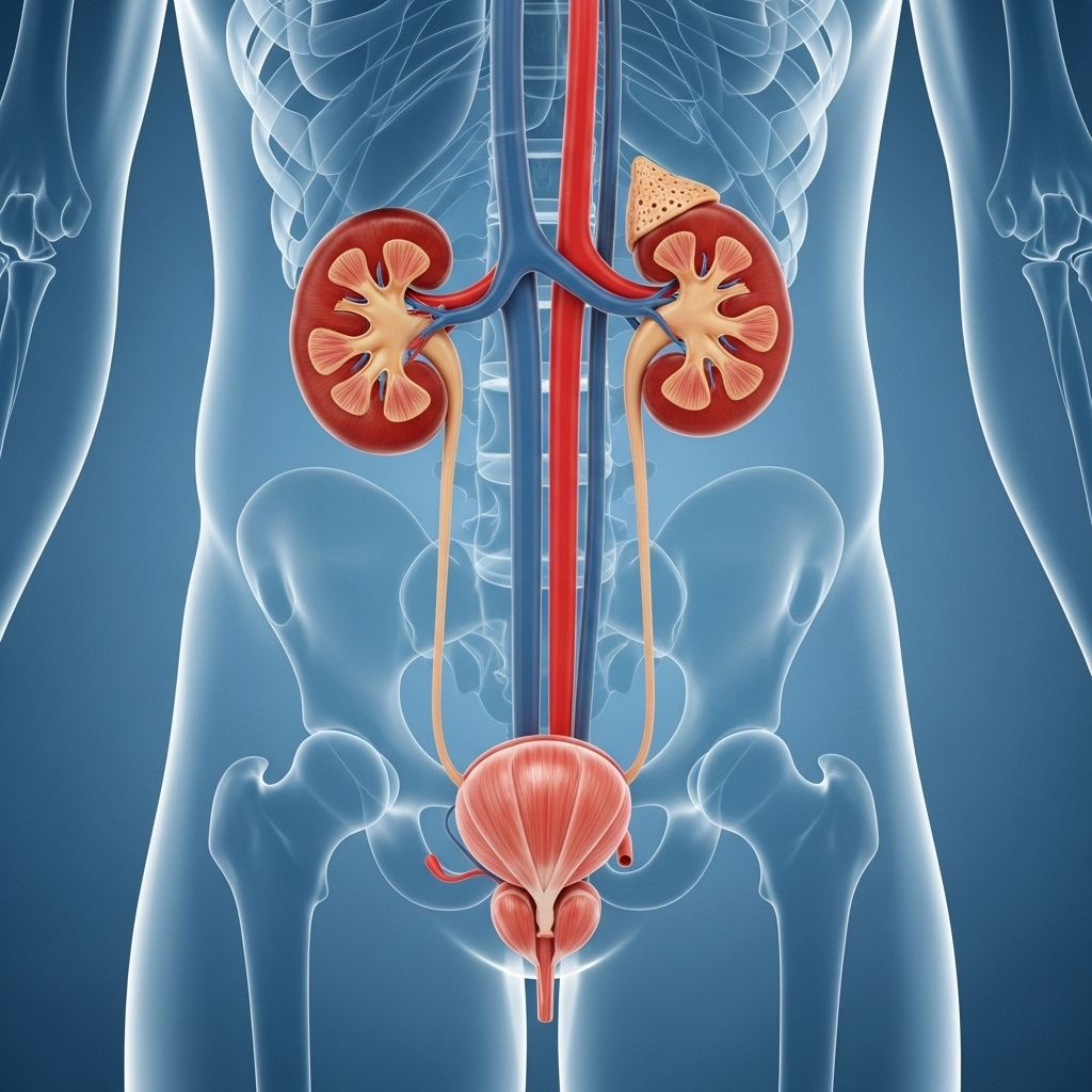
The urinary system—sometimes called the renal system or urinary tract—is a network of organs and ducts working together to remove metabolic waste, maintain fluid and electrolyte balance, and regulate blood pressure. This sophisticated system ensures the proper elimination of waste products while maintaining homeostasis. The urinary system includes four main components: the kidneys, ureters, bladder, and urethra.
Urinary System Overview
- Kidneys: Filter blood to remove waste and excess substances, forming urine.
- Ureters: Carry urine from the kidneys to the bladder.
- Bladder: Stores urine until it can be expelled.
- Urethra: Transports urine from the bladder out of the body during urination.
The Kidneys: Structure and Function
Kidneys are two bean-shaped organs located retroperitoneally on either side of the spine, just below the ribcage. Each kidney is roughly the size of a fist and is protected by surrounding muscle, fat tissue, and the lower ribs.
- The outer renal cortex filters blood to start the process of urine formation.
- The inner renal medulla contains structures called renal pyramids that process the filtered fluid.
- The renal hilum is a concave area where blood vessels, nerves, lymphatics, and the ureter enter and exit the kidney.
Blood enters the kidneys through the renal arteries and leaves via the renal veins. Each kidney receives about 25% of the blood pumped by the heart with every beat, allowing efficient filtration.
Major Functions of the Kidneys
- Filter nitrogenous wastes from the blood.
- Regulate fluid and electrolyte balance.
- Release hormones such as erythropoietin.
- Control blood pressure through renin production.
- Activate vitamin D.
Internal Kidney Structures
- Renal Cortex: Outer layer, home to glomeruli for blood filtration.
- Renal Medulla: Inner layer, containing tubules and the loop of Henle, which concentrates urine.
- Renal Pelvis: Funnel-shaped cavity that collects urine before transfer to the ureter.
- Major and Minor Calyces: Branching structures that funnel urine toward the renal pelvis.
Comparison Table: Kidney Functions
| Function | Description | Location |
|---|---|---|
| Filtration | Removal of waste and excess substances from blood | Renal cortex (glomeruli) |
| Reabsorption | Return of essential substances to blood | Renal tubules |
| Secretion | Active transport of substances into urine | Renal tubules |
| Urine concentration | Removal of water to concentrate urine | Loop of Henle, collecting ducts |
| Hormonal regulation | Secretion of renin, erythropoietin, and activation of vitamin D | Juxtaglomerular apparatus and parenchymal cells |
Ureters: Transporting Urine
The ureters are muscular ducts about 30 centimeters long, running from the renal pelvis of each kidney to the urinary bladder. Retroperitoneal in position, they are anchored between the peritoneum and the posterior abdominal wall by an adventitial layer of collagen and fat.
- Urine is propelled through the ureters by peristaltic contractions of smooth muscle—active waves that prevent passive drainage and work against gravity.
- The inner lining consists of transitional epithelium for flexibility and protection.
- Goblet cells secrete mucus to protect the ureter from the acidity of urine.
The ureters enter the bladder obliquely, which creates a valve-like effect to prevent backflow of urine from the bladder (vesicoureteral reflux).
The Bladder: Storage and Function
The urinary bladder is a hollow, muscular sac situated in the pelvic cavity. Its primary role is to collect and hold urine delivered by the ureters until urination occurs.
- Located anterior to the uterus in females and posterior to the pubic bone and anterior to the rectum in both sexes.
- In males, the prostate gland is found just below the bladder.
- The bladder is partially retroperitoneal; when distended, its dome projects into the abdomen.
A key anatomical feature is the trigone: a triangular-shaped area at the base of the bladder defined by the two ureteral openings and the internal urethral opening. This region is clinically significant as infections often begin here.
Bladder Muscle Layers
- Detrusor muscle: Three layers of smooth muscle that contract to expel urine.
- Transitional epithelial lining: Provides stretch and protection.
During late pregnancy, bladder capacity may be reduced due to compression by the expanding uterus, leading to increased urinary frequency.
The Urethra: Urinary Excretion
The urethra is the final passage for urine, carrying it from the bladder to the exterior body. It is the only part of the urinary tract with significant anatomical differences between males and females.
Male vs. Female Urethra Comparison
| Feature | Male Urethra | Female Urethra |
|---|---|---|
| Length | ~20 cm | ~4 cm |
| Function | Transports urine & semen | Transports urine only |
| Epithelium | Transitional, pseudo-stratified columnar, and stratified squamous | Transitional and stratified squamous |
| Location | Passes through prostate and penis | Opens anterior to vaginal opening |
In both genders, the proximal urethra is lined with transitional epithelium, while the distal portion is stratified squamous epithelium. In men, a segment is lined with pseudostratified columnar cells. The process of urination is regulated by two sphincters:
- Internal urinary sphincter: Involuntary, composed of smooth muscle, controlled by the autonomic nervous system.
- External urinary sphincter: Voluntary, made of skeletal muscle, allowing conscious control over urination.
The urethral opening, location, and length make females more susceptible to urinary tract infections due to proximity to the rectum and a shorter passage for bacteria to travel.
Urine Formation and Elimination
Urine formation is essential for homeostasis, removing excess water, electrolytes, and metabolic wastes. The kidneys filter the blood, forming a filtrate that passes through a sequence of modifications in the renal tubules, finally becoming urine and exiting via the ureters, bladder, and urethra.
- Filtration: Takes place in glomeruli, removing small molecules and water from the blood.
- Reabsorption: Valuable nutrients and water are returned to the circulation.
- Secretion: Additional waste substances are actively transported into the filtrate.
The coordinated action of these processes maintains pH balance, blood volume, and correct electrolyte concentrations.
Clinical Significance & Common Urinary System Issues
The urinary system is subject to several health concerns:
- Urinary tract infections (UTIs): More frequent in women due to urethral anatomy.
- Kidney stones: Mineral deposits forming in the renal pelvis, potentially obstructing urine flow.
- Incontinence: Inability to control urination, related to sphincter dysfunction.
- Renal failure: Inadequate filtration and waste removal, resulting in dangerous toxicity.
- Bladder disorders: Include cystitis, overactive bladder, and bladder cancer.
Early detection and management of urinary system problems are crucial for overall well-being.
Maintaining Urinary System Health
- Stay hydrated by drinking adequate water.
- Practice proper hygiene to reduce infection risk.
- Avoid excessive salt and protein intake to minimize kidney stone risk.
- Monitor urinary habits and seek medical advice for changes.
- Manage underlying health conditions such as diabetes and hypertension.
Frequently Asked Questions (FAQs)
Q: What are the main organs of the urinary system?
A: The main organs are the kidneys, ureters, bladder, and urethra.
Q: How do the kidneys filter blood?
A: Kidneys filter blood through specialized structures called glomeruli, removing waste, excess water, and electrolytes to produce urine.
Q: Why are females more prone to UTIs?
A: The female urethra is shorter and closer to the rectum, increasing the risk of bacterial migration and infection.
Q: What is the function of the bladder?
A: The bladder stores urine before it is eliminated through urination.
Q: How does urine travel from the kidneys to the bladder?
A: Urine passes through the ureters via wave-like contractions called peristalsis.
Q: What actions support urinary system health?
A: Sufficient hydration, proper hygiene, dietary moderation, and routine medical checkups all help maintain urinary health.
Summary Table: Key Points About the Urinary System
| Component | Main Function | Key Details |
|---|---|---|
| Kidneys | Filter blood, produce urine | Bean-shaped, retroperitoneal, regulates fluid/electrolytes |
| Ureters | Transport urine to bladder | Muscular tubes, peristalsis, ~30 cm length |
| Bladder | Store and expel urine | Detrusor muscle, trigone, variable capacity |
| Urethra | Eliminate urine from body | Length and function differs between sexes, two sphincters |
Understanding the anatomy and function of the urinary system helps foster better health practices and prompt attention to potential medical issues. This system is vital to overall wellness, highlighting the importance of regular medical evaluation and healthy lifestyle choices for long-term urinary health.
References
Read full bio of medha deb


