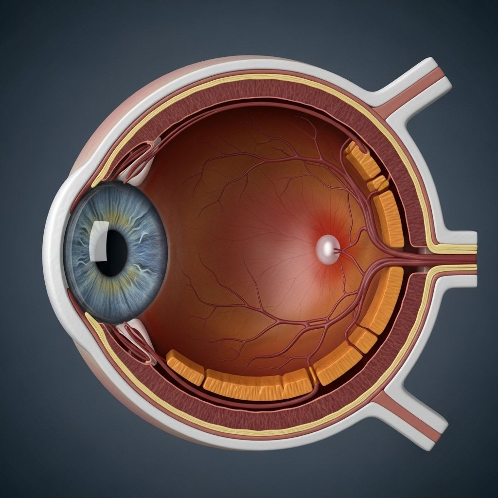
Anatomy of the Eye: Structure, Function, and Common Conditions
Explore the intricate structures and vital functions of the human eye, and how they work together to support vision and overall eye health.

The human eye is a marvel of biological engineering, designed to collect light, process visual information, and deliver clear images to the brain. Understanding the complex anatomy of the eye provides crucial insights into how we see the world and what can go wrong when disease or injury affects vision. This article explores the main components of the eye, their functions, and common disorders that may affect them.
Overview of Eye Anatomy
The eye is a highly specialized sensory organ, tasked with capturing light and translating it into electrical signals interpreted by the brain as vision. The globe of the eye is composed of multiple layers and structures, each with a unique role. Collectively, these parts ensure accurate focusing, image detection, and protection from external damage.
- Cornea: The clear outermost layer that helps focus incoming light.
- Iris: Colored part of the eye that regulates light entering the pupil.
- Pupil: Central opening in the iris that adjusts to control the amount of light reaching the lens.
- Lens: Transparent structure that fine-tunes focus onto the retina.
- Retina: Light-sensitive layer at the back of the eye that triggers nerve impulses.
- Optic nerve: Pathway that carries visual signals to the brain.
Main Structures of the Eye and Their Functions
External Eye Structures
- Eyelids (Palpebrae): Protect the eye from injury, dust, and excessive light; help spread tears evenly across the surface.
- Eyelashes: Trap debris and prevent particles from entering the eye.
- Conjunctiva: Thin, transparent tissue that lines the inside of the eyelids and covers the white of the eye (sclera); protects against infection.
- Lacrimal Gland: Produces tears to lubricate and protect the eye.
Anterior Segment
| Part | Description | Function |
|---|---|---|
| Cornea | Clear, dome-shaped surface that covers the front of the eye | Focuses light as it enters the eye; provides most of the eye’s optical power |
| Aqueous Humor | Clear fluid filling the space between cornea and lens | Nourishes cornea and lens; maintains intraocular pressure |
| Iris | Colored muscular ring | Adjusts pupil size to control light entry |
| Pupil | Opening in center of iris | Regulates amount of light entering the lens |
| Lens | Transparent, biconvex structure | Fine-tunes focus by changing shape (accommodation) |
| Ciliary Body | Ring of muscle and connective tissue | Alters lens shape for near/far focus; produces aqueous humor |
| Sclera | Opaque ‘white’ of the eye | Maintains eye shape; protects internal structures |
Posterior Segment
| Part | Description | Function |
|---|---|---|
| Vitreous Humor | Clear, gel-like substance filling the interior behind the lens | Maintains eye shape; transmits light to the retina |
| Retina | Thin, neural layer lining back wall of the eye | Contains photoreceptors (rods and cones) for vision |
| Macula | Small central area of retina | Provides sharp, detailed central vision |
| Fovea | Center of the macula | Area of highest visual acuity; densely packed with cones |
| Choroid | Vascular layer between retina and sclera | Supplies oxygen and nutrients to retina |
| Optic Disc | Site where optic nerve exits the eye | Known as the ‘blind spot’; no photoreceptors |
| Optic Nerve | Bundle of nerve fibers | Transmits visual information to the brain |
How the Eye Works: The Visual Pathway
Vision begins when light rays enter the eye through the cornea, which bends and focuses the incoming light. The light then passes through the pupil, the opening in the iris that adjusts in size based on lighting conditions. The lens further refines the focus by changing its curvature, a process known as accommodation.
After traversing the lens, the light passes through the vitreous humor and finally lands on the retina. Specialized photoreceptor cells—rods (for low-light and peripheral vision) and cones (for color and detailed central vision)—convert the light into electrical signals. These signals are processed through several layers of retinal neurons and transmitted via the optic nerve to the brain’s visual cortex, where they are interpreted as images.
Key Parts of the Retina
- Rods: Sensitive to low light, important for night and peripheral vision.
- Cones: Detect color and detail, concentrated in the fovea.
- Macula: Central area with highest density of cones for acute vision.
- Retinal Pigment Epithelium: Supports photoreceptor health and absorbs excess light.
Protective Mechanisms of the Eye
The eye employs several defensive systems to maintain its integrity and function:
- Blink Reflex: Shields against foreign objects and bright light.
- Tear Production: Lubricates, cleans, and delivers nutrients to the eye surface.
- Sclera and Cornea: Form durable barriers against injury and infection.
- Immune Cells: Present in the conjunctiva and other layers to fend off pathogens.
Common Eye Conditions Related to Anatomy
Understanding eye anatomy aids in recognizing common eye conditions, many of which are related to the malfunction or injury of a particular structure.
- Conjunctivitis (Pink Eye): Inflammation of the conjunctiva, leading to redness and irritation.
- Cataracts: Clouding of the lens, causing blurry vision and glare.
- Glaucoma: Damage to the optic nerve often associated with elevated intraocular pressure, potentially leading to vision loss.
- Macular Degeneration: Degeneration of the macula, impairing central vision.
- Retinal Detachment: Separation of the retina from underlying tissue, leading to vision loss if untreated.
- Diabetic Retinopathy: Damage to retinal blood vessels from chronic high blood sugar.
- Dry Eye Syndrome: Insufficient tear production or poor tear quality causing discomfort and visual disturbance.
Maintaining Eye Health
Several lifestyle factors play a significant role in protecting the eyes and supporting visual health throughout life:
- Regular eye examinations help detect problems early, even before symptoms arise.
- Proper nutrition (including vitamins A, C, E, and zinc, and omega-3 fatty acids) supports retinal and overall eye health.
- Wearing protective eyewear in dangerous or sporting environments prevents injury.
- Minimizing screen time and taking frequent breaks can reduce eye strain.
- Managing chronic conditions such as diabetes greatly reduces the risk for certain eye diseases.
- UV protection (sunglasses that block UVA/UVB light) is essential to prevent damage from the sun’s rays.
Frequently Asked Questions (FAQs) About Eye Anatomy
Q: What gives the eye its color?
A: The color of the eye is determined by the amount and type of pigmentation present in the iris.
Q: Why do some people need glasses or contact lenses?
A: Refractive errors occur when the shape of the eye prevents light from focusing directly on the retina, leading to nearsightedness, farsightedness, or astigmatism. Glasses or contact lenses correct these errors by adjusting the way light rays enter the eye.
Q: What is the blind spot?
A: The blind spot, or optic disc, is where the optic nerve exits the eyeball. Since there are no rods or cones in this area, it cannot detect light, resulting in a natural blind spot in the visual field.
Q: How does the eye adjust to see objects at different distances?
A: The ciliary muscles change the shape of the lens through accommodation, allowing for clear focus on objects both near and far.
Q: What is the function of tears?
A: Tears moisturize the surface of the eye, provide nutrients, contain protective enzymes against microbes, and wash away irritants.
Glossary of Key Eye Anatomy Terms
| Term | Definition |
|---|---|
| Cornea | Transparent front part of the eye |
| Iris | Colored part of the eye; regulates pupil size |
| Pupil | Opening in the iris; allows light to enter |
| Lens | Adjusts focus for near and distant vision |
| Retina | Light-sensitive tissue lining the back of the eye |
| Macula | Area of retina with highest visual acuity |
| Optic nerve | Pathway to the brain for visual information |
| Sclera | White, protective outer covering of the eye |
| Ciliary body | Controls lens shape and produces aqueous humor |
| Vitreous humor | Gel filling rear segment of the eye |
Quick Facts and Tips for Eye Health
- The cornea provides about two-thirds of the eye’s total focusing power.
- There are about 120 million rods and 6 million cones in the human retina.
- The macula is responsible for the sharp, central vision needed for activities like reading and driving.
- The optic nerve is made of more than a million individual nerve fibers.
- Regular eye exams are essential for early detection of eye diseases such as glaucoma and macular degeneration.
When to See an Eye Specialist
Seek prompt eye care if you experience any of the following:
- Sudden change in vision or loss of vision
- Floaters, flashes, or shadows in vision
- Eye pain or redness lasting more than a day
- Persistent dryness, itching, or discharge
Early intervention often prevents minor problems from becoming serious threats to sight.
References
- https://www.jhuapl.edu/news/news-releases/250116-retinal-selfie
- https://www.nei.nih.gov/about/news-and-events/news/johns-hopkins-medicine-scientists-uncover-molecular-link-between-wet-and-dry-macular-degeneration
- https://optometry.osu.edu/academics/residency-programs/program-descriptions/johns-hopkins-medicine-wilmer-eye-institute
- https://myasco.opted.org/searchEngines/residency_details.aspx?id=345
- https://hub.jhu.edu/2021/10/14/greenberg-center-to-end-blindness/
Read full bio of Sneha Tete


