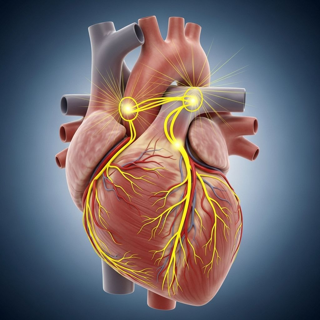
Anatomy and Function of the Heart’s Electrical System
Understanding how the heart’s electrical system controls heartbeat, rhythm, and healthy cardiac function.

The heart, often described as the body’s living engine, relies on a precisely organized electrical system to sustain life. This built-in wiring ensures that the heart’s muscular pump beats in a steady rhythm, efficiently distributing blood throughout the body. The complexity and reliability of the heart’s electrical circuit are remarkable—and essential for both health and survival.
Overview: The Heart’s Electrical Network
At its core, the heart is a muscular pump. Unlike any other muscle, however, its contractions are tightly coordinated by a network of specialized cells capable of initiating and conducting electrical impulses. These impulses instruct the heart’s chambers to contract in a perfectly timed sequence, allowing for the continuous and rhythmic flow of blood.
- The heart’s rhythm is generated and maintained by an electrical conduction system.
- This system ensures four heart chambers (two atria, two ventricles) contract in the correct order.
- Heart rate (normally 60–100 beats per minute in adults) is determined by electrical signals.
Anatomy of the Heart’s Electrical System
The heart’s electrical system consists of specialized cells and structures embedded within the heart muscle. Each structure plays a distinct role in the propagation of the electrical impulse that triggers myocardial contraction.
| Structure | Location | Function |
|---|---|---|
| Sinoatrial node (SA node) | Right atrium, near superior vena cava | Primary pacemaker; initiates each heartbeat |
| Atrioventricular node (AV node) | Boundary between atria and ventricles | Delays signal briefly to allow atria to contract |
| Bundle of His | Upper part of interventricular septum | Transmits impulse from AV node to bundle branches |
| Right and left bundle branches | Along interventricular septum | Conduct impulses toward ventricles |
| Purkinje fibers | Spread throughout ventricular walls | Quickly distribute electrical impulse within ventricles |
This interconnected system ensures that electrical impulses are delivered efficiently and systematically, resulting in coordinated heart muscle contractions.
The Conduction Pathway: How an Electrical Pulse Travels
Each heartbeat is driven by a wave of electrical activity that follows a specific, repeating pathway:
- Generation at the SA Node: The sinoatrial (SA) node—located in the upper right atrium—acts as the heart’s natural pacemaker, producing electrical impulses 60–100 times per minute in a healthy adult. This impulse sets the pace and rhythm of the heartbeat.
- Atrial Contraction: The impulse spreads through the right and left atria, causing these upper chambers to contract. Blood is thereby pushed from the atria into the ventricles.
- Delay at the AV Node: Electrical signals then reach the atrioventricular (AV) node, located at the junction between the atria and ventricles. Here, the signal is briefly delayed. This pause allows the ventricles time to fill completely with blood before contracting.
- Impulse Down the Bundle of His: After the delay, the impulse travels rapidly down the bundle of His, penetrating the interventricular septum.
- Transmission Through Bundle Branches: The bundle of His splits into the right and left bundle branches, which direct the impulse toward the heart’s apex and the ventricles’ muscle walls.
- Purkinje Fibers Activate Ventricles: Finally, Purkinje fibers disperse the electrical signal throughout the ventricles, causing a powerful and synchronized contraction. This propels blood out of the heart—into the lungs from the right ventricle, and the rest of the body from the left ventricle.
Key Point: The well-timed progression and delay between atrial and ventricular contraction maximize the efficiency of blood flow.
How the Heartbeat Cycle Works
The cardiac cycle is the sequence of events occurring from the start of one heartbeat to the beginning of the next, orchestrated entirely by the electrical system:
- Atrial contraction occurs first, triggered by the SA node’s firing.
- Blood is pushed into the relaxed ventricles.
- The impulse reaches the AV node, is delayed, then passed on to the ventricles.
- Ventricular contraction follows—blood is forced into the arteries.
- The myocardium (heart muscle) relaxes—atria fill with blood in preparation for the next cycle.
In a typical adult at rest, this cycle repeats 60 to 100 times per minute, depending on age and overall health.
Regulation of Heart Rate and Rhythm
The heart’s electrical activity is influenced by internal and external factors. Key regulators include:
- Autonomic nervous system (ANS): The sympathetic branch accelerates heart rate, while the parasympathetic branch slows it down.
- Hormones: Catecholamines (like adrenaline) from the adrenal glands increase rate and contractility during stress or excitement.
- Electrolyte levels: Potassium, calcium, and sodium balance is essential for proper electrical conduction.
- Age: Heart rate generally slows with increasing age.
- Body condition: Exercise, temperature, medication, illness, and metabolic state can also affect the heart’s electrical pace.
The heart’s ability to automatically generate electrical impulses, known as automaticity, allows it to beat independently of direct neural input, yet remain responsive to wider physiological demands.
Common Disorders of the Heart’s Electrical System
While the electrical system typically functions seamlessly, disruptions can cause an array of cardiac arrhythmias or conduction problems:
- Sinus node dysfunction: An underactive SA node (sick sinus syndrome) can cause the heart to beat too slowly.
- AV node block: Delay or blockage in the AV node (heart block) interrupts the relay of signals from atria to ventricles.
- Bundle branch block: Delay or failure in one branch slows ventricular activation, affecting coordination.
- Atrial fibrillation: Disorganized electrical activity in the atria leads to rapid, irregular heartbeats.
- Ventricular tachycardia/fibrillation: Abnormal, very fast rhythms in the ventricles, especially dangerous and may prevent effective pumping of blood.
Symptoms of disrupted cardiac conduction can range from palpitations and fatigue to fainting and, in extreme cases, sudden cardiac arrest.
Diagnosing Electrical System Problems
Identifying abnormalities within the heart’s electrical system usually begins with an evaluation of symptoms and a physical exam. Diagnostic tests may include:
- Electrocardiogram (ECG/EKG): Records the heart’s electrical activity over time, revealing rhythm or conduction abnormalities.
- Holter monitor: A portable ECG device worn for 24-48 hours to track ongoing electrical activity.
- Event recorder: Activated by a patient to record symptoms as they occur over weeks.
- Electrophysiology studies: Intracardiac catheters and electrodes map the heart’s conduction in detail, helping guide therapeutic interventions.
Treatment of Conduction Disorders
Treatment depends on the specific disorder and its severity:
- Medications: Antiarrhythmics, beta-blockers, and calcium channel blockers help stabilize or control heart rhythms.
- Implantable devices: Pacemakers restore normal rhythm in cases of slow heart rate or blocks by providing artificial electrical stimulation.
- Defibrillators: Implantable cardioverter-defibrillators (ICDs) monitor heart rhythm continuously and deliver life-saving shocks for dangerous ventricular arrhythmias.
- Catheter ablation: Uses radiofrequency energy to selectively destroy small areas of abnormal tissue that cause arrhythmias.
- Lifestyle management: Controlling risk factors—such as hypertension, sleep apnea, or drug/alcohol use—can improve outcomes.
Preserving the Heart’s Electrical Health
While some electrical disorders are inherited or age-related, many risk factors can be managed:
- Control blood pressure and cholesterol through healthy lifestyle choices.
- Avoid tobacco and limit alcohol intake.
- Maintain a healthy weight and exercise regularly.
- Manage underlying medical conditions such as diabetes and sleep apnea.
- Follow physician guidance to monitor or treat arrhythmias and heart disease.
Frequently Asked Questions (FAQs)
Q: What is the SA node, and why is it called the heart’s pacemaker?
The sinoatrial node (SA node) is a group of specialized cells in the upper right atrium. It produces electrical impulses that set the pace and rhythm of the heartbeat, earning it the title “natural pacemaker.”
Q: Why does the AV node delay the electrical signal?
The brief delay at the atrioventricular (AV) node allows the atria to contract and fully empty blood into the ventricles before ventricular contraction begins, ensuring maximum efficiency in blood flow.
Q: What happens if the electrical system malfunctions?
Malfunctions can lead to arrhythmias (irregular rhythms), heart block (delayed or blocked conduction), or other conduction disorders, which may affect the heart’s ability to pump blood effectively and could result in symptoms or health risks.
Q: Can the heart beat without input from the brain?
Yes. The heart’s electrical system (primarily the SA node) is capable of initiating its own impulses independently, though its rate and strength are influenced by the autonomic nervous system.
Q: How are arrhythmias treated?
Treatments for arrhythmias vary and may include medications, pacemakers, implantable defibrillators, ablation procedures, and lifestyle modifications, depending on the underlying disorder, severity, and patient health.
Key Takeaways
- The heart’s electrical system is vital for coordinating the contractions of every heartbeat.
- Specialized nodes and pathways ensure precise timing and direction of electrical signals.
- Arrhythmias and conduction blocks can disrupt this harmony, but diagnosis and modern treatments often restore function and quality of life.
Understanding the anatomy and function of the heart’s electrical circuitry provides critical insight into how the heart maintains its remarkable pace—and what can be done when its rhythm falters.
References
- https://www.ummhealth.org/health-library/anatomy-and-function-of-the-hearts-electrical-system
- https://www.urmc.rochester.edu/encyclopedia/content?contenttypeid=90&contentid=p01762
- https://www.stanfordchildrens.org/en/topic/default?id=anatomy-and-function-of-the-electrical-system-90-P01762
- https://www.youtube.com/watch?v=YWWVSW0AWgw
- https://my.clevelandclinic.org/health/body/21648-heart-conduction-system
- https://myhealth.alberta.ca/Health/pages/conditions.aspx?hwid=te7147abc
- https://my.clevelandclinic.org/health/body/21704-heart
Read full bio of Sneha Tete


