Peering Into Nature: Rob Kesseler’s Artistic Micrographs of Pollen, Seeds, and Fruit
Discover how Rob Kesseler transforms microscopic plant structures into mesmerizing art, merging scientific inquiry with creative vision.

Rob Kesseler: Where Art and Science Meet Under the Microscope
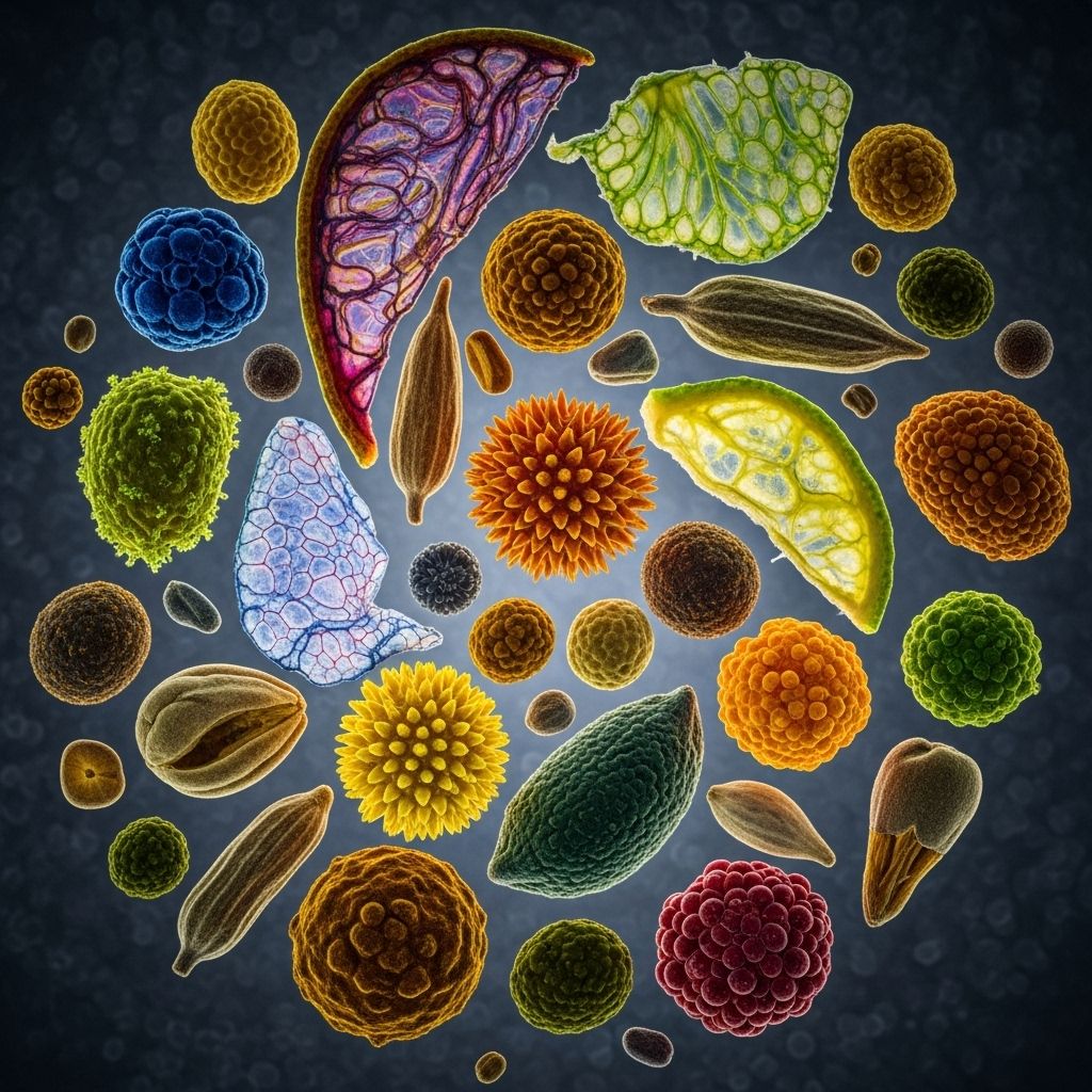
Rob Kesseler, an acclaimed British artist and researcher, has pioneered a unique fusion of art and science—producing mesmerizing photographic micrographs of pollen, seeds, and fruit. These images, achieved through scanning electron microscopy (SEM), showcase intricate plant structures typically hidden from the naked eye, their patterns and colors meticulously enhanced and hand-colored to accentuate both scientific detail and artistic wonder.
Kesseler, Chair of Arts, Design & Science at Central Saint Martins, and a fellow of societies such as the Linnean Society and the Royal Microscopical Society, is at the forefront of interdisciplinary collaboration. His award-winning publications on pollen, seeds, and fruit, alongside high-profile projects with scientists, elevate microscopic plant images to the realm of fine art.
Below, we take a comprehensive look at how Kesseler creates, colors, and contextualizes these captivating works—revealing not only the astonishing complexity of nature’s smallest features but also the innovative ways that art and science can intersect.
The Genesis of a Vision: Childhood Microscope to Global Impact
The seeds for Kesseler’s creative journey were sown in childhood when his father gifted him a microscope—a moment he describes as transformative. This early exposure fostered a sense of curiosity about the hidden worlds within ordinary objects. In Kesseler’s own words, the microscope granted him “a second vision” and a heightened awareness of another dimension composed of forms, colors, and patterns beyond normal sight.
His lifelong passion for observation and research underpins his art. Kesseler’s work is a testimony to the profound inspiration found in the natural world, and how scientific tools can expand artistic horizons.
The Artistic Technique: Scanning Electron Microscopy and Hand-Coloring
To unveil the elaborate architecture of plant cells, pollen grains, and seeds, Rob Kesseler employs scanning electron microscopy (SEM)—a technique that enables the visualization of surfaces at magnifications up to tens of thousands of times, far surpassing what traditional light microscopes can achieve.
After capturing stunningly detailed grayscale images, Kesseler applies color by hand, layering pigment digitally or manually to accentuate forms and evoke emotional responses. This process is informed by both scientific observation and his artistic intuition. The interplay between monochrome SEM imagery and vivid, lifelike coloration transforms data into visual poetry.
- SEM reveals ultra-fine surface textures—grooves, spikes, and lattices on tiny grain and seed surfaces.
- Color is added post-capture, letting Kesseler highlight key structures and direct the viewer’s gaze.
- This marriage of precision and subjective enhancement draws viewers into a world at once objective and interpretive.
Exploring Pollen: Nature’s Microscopic Architecture
Pollen grains, vital in plant reproduction, stand as some of nature’s most intricate microstructures. Under Kesseler’s lens, their alien-like forms—ranging from spiky spheres to honeycombed or netted surfaces—take on both aesthetic and educational significance.
Pollen grains feature highly specific patterns that often hold clues about a species’ evolutionary adaptations. Colorizing these, Kesseler not only brings clarity to their structure but also highlights the evolutionary artistry of the natural world.
| Pollen Species | Microscopic Features | Artistic Enhancement |
|---|---|---|
| Lilium (Lily) | Large, ornamented grains with distinct exine patterns | Bright color contrasts to highlight exine ridges |
| Ranunculus | Rounded grains with a spongy texture | Soft lighting and color overlays emphasize lush surfaces |
| Wheat | Smooth, aerodynamic shape; modest ornamentation | Subtle color washes illuminate structure |
Kesseler’s pollen micrographs appear in scientific slide kits as well as art books, demonstrating their dual value for botanists and art lovers alike.
For example, digitally enhanced images from microscope slide kits show three varieties of pollen at high magnification (400x), each revealing distinct reproductive adaptations.
Seeds and Fruit: Unveiling Diversity at the Cellular Level
Seeds represent the culmination of the plant reproductive cycle, yet their form and texture often remain invisible until subjected to extreme magnification. Kesseler’s photographs illuminate these hidden worlds, showing each seed’s protective coat, sculptural geometry, and adaptation for dispersal.
The surface of fruit, such as the Prunus persica (peach), displays a universe of miniature features—pits, ridges, and vessels—that serve ecological functions and provide visual intrigue.
Stacked macro photography and SEM imaging reveal:
- Pear sections with visible cellular grids, appearing almost crystalline under the microscope.
- Corn seeds with layered outer coatings that protect nutritional stores.
- Capsella embryos with cotyledons laid out like blueprints for future growth.
Kesseler’s use of color not only draws attention to these features but also humanizes botanical forms that could otherwise seem alien.
The Intersection of Art and Science: Morphogenetic Synthesis
Rob Kesseler refers to his process as a “morphogenetic synthesis”—a productive overlap between two expansive cultures: art and science. His micrographs are both analytical tools for researchers and objects of aesthetic contemplation. This approach reflects a renewed era in which artists and scientists collaborate, moving beyond the century-old separation of disciplines.
Kesseler’s work with leading scientists, including collaborations at the Royal Botanical Gardens in Kew, Oxford Instruments, and the BBC, produces not just remarkable images but also fosters new ways to communicate research to the public.
His perspective is that “after a century of separation, artists and scientists are again working together, sharing ideas that reflect our age.” This synergy is visible in his imagery—art used to attract attention and science as a source of endless curiosity.
Broadening Scientific Understanding Through Visual Art
Microscopic art such as Kesseler’s is far more than a visual curiosity; it’s a catalyst for scientific engagement. Pioneers at research institutions, including collaborations with seed conservation labs, are using microscopic photography to document, preserve, and study biodiversity—especially among endangered plant species.
For example, Hawaiian flora researchers found that traditional photography struggled to capture tiny seed detail. The adoption of stereomicroscopy and digital imaging enabled catalogues of hundreds of rare species, contributing valuable data for conservation.
- Microscope micrographs can help identify, catalogue, and conserve threatened plant species.
- High-resolution imagery aids botanists in classifying plants and understanding reproductive ecology.
- Making these images publicly available encourages citizen science and global education.
Nature’s Invisible Beauty: Patterns, Colors, and Impact
The patterns revealed through microscope photography—spikes, honeycomb latticework, and sculpted hills—may appear alien yet are patterned by evolutionary pressures for survival and dispersal. Kesseler’s color palette, inspired by long hours of research and observation, is carefully chosen to both attract attention and remain true to nature.
These works underscore the interconnectedness of form and function. A grain’s ridges may ensure it sticks to pollinators; seed coats may resist moisture or predation; fruit surfaces might guide dispersal. Artistic interpretation brings these biological narratives into focus for broader audiences.
Recognition and Influence: Books, Collaborations, and Public Engagement
Rob Kesseler’s micrographs have appeared globally in scientific books, fine art exhibitions, and media collaborations. His publications on pollen, seeds, and fruit—produced with Papadakis—have won critical acclaim for bridging the divide between scientific knowledge and aesthetic appreciation.
Recent joint projects with journalists and botanists explore climate change and its impact on plant diversity, making microscopic art a tool for raising ecological awareness.
- Chair of Arts, Design and Science at Central Saint Martins, University of the Arts London.
- Fellowships with the Linnean Society, Royal Society of Arts, Royal Microscopical Society.
- Collaborative projects with global research institutions and conservation labs.
The Future: Art, Science, and New Ways of Seeing
As interdisciplinary collaboration intensifies, projects like Kesseler’s point toward a future where art and science are complementary rather than compartmentalized. Increasingly, both fields recognize the importance of creative visualizations in communicating complex data, inspiring innovation, and fostering public engagement.
Kesseler hopes “we are entering a new age” of idea-sharing, where the detailed study of form and color can reveal not only the biology of plants but the broader patterns connecting all living things.
Frequently Asked Questions (FAQ)
Q: What is scanning electron microscopy (SEM)?
A: SEM is a powerful microscopy technique that uses electrons rather than light to scan surfaces at extremely high magnifications, revealing minute details of structure such as the surface of pollen grains and seeds.
Q: How does Rob Kesseler add color to his micrographs?
A: Kesseler manually and digitally enhances SEM images by hand-coloring them—a process that requires both scientific observation and artistic intuition to illuminate structural features and attract attention.
Q: Why is microscope photography important for plant science?
A: Microscopy allows scientists to study the intricate forms and adaptations of pollen, seeds, and fruit, which can be crucial for taxonomy, conservation, and understanding plant evolution and ecology.
Q: Are Kesseler’s photographs used in research as well as art?
A: Yes—his colored micrographs appear in scientific slide kits, research publications, and are used to support plant identification and classification, as well as engaging general audiences in art exhibits.
Q: What impact do these images have on public understanding?
A: By revealing hidden patterns and structures, the images foster appreciation for biodiversity and highlight the importance of plant life in ecological health, conservation, and climate awareness.
Conclusion: Nature’s Details Reimagined
Rob Kesseler’s artistic microscope photography allows viewers to peer into an unseen dimension of nature—making the invisible visible, and redefining our relationship with the microscopic beauty of everyday plants. His work not only enriches scientific inquiry but ignites the imagination and elevates the conversation between art and science.
Through his lens, pollen, seeds, and fruit become ambassadors for their species, drawing attention to their survival and adaptation in a changing world, and reminding us of the elegant complexity that underpins all life.
References
- https://www.thisiscolossal.com/2019/12/rob-kesseler-pollen-photographs/
- https://www.shutterstock.com/search/microscopic-pollen?image_type=photo&page=2
- https://www.microscopeworld.com/t-fruit_flower_microscope_slides.aspx
- https://saveplants.org/video/revealing-the-details-of-hawaiian-seeds-through-microscope-photography/
- https://mymodernmet.com/rob-kesseler-photographs/
Read full bio of Sneha Tete


